Published online Aug 27, 2016. doi: 10.4240/wjgs.v8.i8.590
Peer-review started: April 20, 2016
First decision: June 6, 2016
Revised: June 23, 2016
Accepted: July 11, 2016
Article in press: July 13, 2016
Published online: August 27, 2016
Processing time: 128 Days and 8.6 Hours
To compare the 3 main techniques of temporary closure of the abdominal cavity, vacuum assisted closure (vacuum-assisted closure therapy - VAC), Bogota bag and Barker technique, in damage control surgery.
After systematic review of the literature, 33 articles were selected to compare the efficiency of the three procedures. Criteria such as cost, infections, capacity of reconstruction of the abdominal wall, diseases associated with the technique, among others were analyzed.
The Bogota bag and Barker techniques present as advantage the availability of material and low cost, what is not observed in the VAC procedure. The VAC technique is the most efficient, not only because it reduces the tension on the boarders of the lesion, but also removes stagnant fluids and debris and acts at cellular level increasing cell proliferation and division. Bogota bag presents the higher rates of skin laceration and evisceration, greater need for a stent for draining fluids and wash-ups, higher rates of intestinal adhesion to the abdominal wall. The Barker technique presents lack of efficiency in closing the abdominal wall and difficulty on maintaining pressure on the dressing. The VAC dressing can generate irritation and dermatitis when the drape is applied, in addition to pain, infection and bleeding, as well as toxic shock syndrome, anaerobic sepsis and thrombosis.
The VAC technique, showed to be superior allowing a better control of liquid on the third space, avoiding complications such as fistula with small mortality, low infection rate, and easier capability on primary closure of the abdominal cavity.
Core tip: The authors reviewed several manuscripts published in the last 20 years evaluating the management of the open abdomen. Several techniques have been described and the most popular ones were analyzed. The vacuum assisted closure (vacuum-assisted closure therapy), Bogota bag and Barker technique are currently the most used. The authors evaluate, for each technique, efficiency, complications, mortality and wound control.
- Citation: Ribeiro Junior MAF, Barros EA, de Carvalho SM, Nascimento VP, Cruvinel Neto J, Fonseca AZ. Open abdomen in gastrointestinal surgery: Which technique is the best for temporary closure during damage control? World J Gastrointest Surg 2016; 8(8): 590-597
- URL: https://www.wjgnet.com/1948-9366/full/v8/i8/590.htm
- DOI: https://dx.doi.org/10.4240/wjgs.v8.i8.590
Throughout history surgical principles were based on anatomical repairs with the goal to make primary and definite organic repair. In the last decade, a greater importance has been given in repairing physiological features of the surgical patient. This resulted in the concept of damage control surgery with special emphasis on the need for open abdominal maintenance (laparotomy)[1].
Laparostomy comprises a surgical approach where the abdominal wall is not sutured by plans and left open, allowing a regular inspection and drainage of intracavitary content[2,3]. Its usage is indicated in cases of abdominal trauma, damage control surgery, sepsis of abdominal origin, severe acute pancreatitis, the need for new laparotomy, hemodynamic instability, retroperitoneal hematomas and necrotizing fasciitis, allowing a continuous evaluation of intra-abdominal pressure in order to avoid the development of abdominal compartment syndrome[2,4-7].
According to the consensus carried out by the World Society of the Abdominal Compartment Syndrome (WSCAS - 2004), abdominal compartment syndrome is defined as a condition where organ dysfunction is caused by sustained intra-abdominal hypertension above 20 mmHg. However, it is known that values below this (> 12 mmHg) will also cause organ dysfunction, but to a lesser degree. Abdominal hypertension is divided in degrees[2]: (1) 12-15 mmHg; (2) 16-20 mmHg; (3) 21-25 mmHg; and (4) > 25 mmHg.
In 2013, the WSACS concluded that intra-abdominal pressure must be measured in patients who have at least two risk factors for hypertension, which are correlated with the pathology that triggered the abdominal injury. This pressure must be measured every 4-6 h, using preferentially the bladder and in cases of contraindication, other via as abdominal, rectal and vaginal may also be used[2,5]. The compartmental syndrome may also be classified according to their etiology (Table 1)[5].
| Primary | Secondary |
| Abdominopelvic causes[5]: | Extra-abdominal causes[5]: |
| Complex abdominal trauma[5] | Sepsis[5] |
| Ruptured Aneurysm aorta[5] | Acidosis (pH < 7.2)[5] |
| Hemoperitoneum[5] | Hypothermia[5] |
| Pancreatitis[5] | Politransfused (> 10 IU of packed red blood cells in 24 h)[5] |
| Peritonitis[5] | Coagulopathies (platelets < 55000/mm3 or aPTT > 2x/normal or TAP < 50% - INR > 1.5 )[5] |
| Retroperitoneal bleeding[5] | Great chest, vascular and orthopedic trauma[5] |
| Hepatic transplants[5] | Great burnt[5] |
| Primary closure under tension[5] | Vigorous hydration (> 5 U/24 h)[5] |
Complications arise from the current shock, which leads to the involvement of multiple systems causing renal perfusion decrease, decline in glomerular filtration rate (oliguria and acute renal insufficiency); ischemia of the intestinal wall and its mucosa, liver failure by portal flow decrease, cerebral hypoperfusion, among others[5].
During the period in which the abdomen is open, the aponeurosis can laterally retract and lose the possibility of closure, creating then a ventral incisional hernia. These, with subsequent formation of adhesions make future abdominal surgeries more complicated, increasing morbidity and mortality[8]. This can be avoided by applying temporary closure techniques, such as Bogota bag, Barker technique, vacuum assisted closure therapy (VAC), among others that allow closing the abdominal cavity with less stress[9].
The aim of this review article is to compare the 3 main techniques of temporary abdominal cavity closure, vacuum closure (VAC), Bogota bag and Barker technique, after damage control laparotomy. In this study, we analyzed efficiency, financial costs, risks and benefits for each of the most common available methods.
This paper is a review of manuscripts published in the literature from 1996 to 2016. Search was performed in MEDLINE, PUBMED, Scielo e Lilac’s with the following keywords: VAC technique, Bogota bag and Barker techniques, traumatic abdominal injuries. The papers should have addressed effectiveness of each technique, cost, associated infections, readiness on abdominal wall reconstruction and diseases associated with the technique. Only articles in English and Portuguese and performed in humans were selected. Thirty-three articles met the selection criteria.
The Bogota bag technique presents as advantages (Table 2) the availability to material access, lower cost, fast and easily executed, and not dependent on a great experience from the surgeon. For allowing a great visibility and entrance in the abdominal cavity, it promotes greater control of the septic spots and enables several revisions. Moreover, it allows abdominal decompression, does not cause inflammatory reactions or allergies, being used in several parts of the organism and in any kind of surgery, from traumas to tumor removal. However, it presents higher rates of eviscerations and skin lacerations, implying greater need for abdominal drainage and wash-ups, higher rates of intestinal adhesion to the abdominal wall, difficulty in controlling the third space and leaks. Thus, it requires sterilization of the bag by gas preventing its availability (Table 3).
| Bogota bag | Allows greater number of reviews of the abdominal cavity than the direct synthesis; better control of septic focuses by easy access; abdominal decompression; and functional restoration of the abdominal wall; has low cost; immediate availability; flexibility and high resistance; is not adhered to the tissues; does not cause allergic or inflammatory reactions; has quick and easy installation; can be used in any part of the body; allows the visualization of organs; is used in trauma, cancer surgery, and various abdominal surgeries; is a protector against water loss and heat |
| Barker technique | It is an inexpensive technique; uses material found in the surgical center and easily applicable; has moderate fluid control; allows closure of the abdominal wall with less tension; has low rates of complications |
| Vacuum-assisted closure therapy | It prevents contamination; allows dissection of the wound; protects the wound from external injuries; reduces the interstitial pressure; increases blood flow to the lesion; reduces the expression of matrix metalloproteinases in chronic wounds; promotes wound healing; removes stagnant fluids and debris; increases proliferation and cell division rates; induces granulation tissue formation; and brings greater comfort to the patient with infrequent complications. In selected cases it can be used as an outpatient procedure; allows shorter hospital stay and better quality of life |
| Bogota bag | Use of more drains and rinses; presents a certain risk of eviscerations and difficulty in mobilizing the patient; it can cause skin lacerations; bowel adhesion to abdominal wall; it needs gas sterilization of the bag; there is a difficult control of the third space; leaks under the bag can wet the bed increasing the risk of hypothermia |
| Barker technique | In some cases, it does not allow adequate approach of the abdominal wall; it has moderate fluid control; greater difficulty in detecting complications that occur with bleeding and the maintaining of continuous pressure |
| Vacuum-assisted closure therapy | High cost; it can cause skin irritation due to the use of the adhesive; it can cause pain, infection and bleeding; it can lead to toxic shock syndrome (rare) and can cause thrombosis |
The technique proposed by Barker presents positive factors, such as easy assembly at a low cost, since it uses hospital material already used for other functions (Table 2). Since acquiring these materials is easy, the technique application becomes simple. The closure of the abdominal cavity is done with less stress, preventing complications associated with moderate control of the fluid produced by the body. One disadvantage is the lack of efficiency in approaching the abdominal wall when the injury is extensive and important. There is a deficiency on maintaining pressure, which must be continuous and properly maintained in order to make the organs act in the recovery. In complicated cases with abdominal bleeding, it may take a while to detect this bleeding while draining the fluid (Table 3).
The temporary closure technique by negative pressure, the VAC, among all of them has a more effective action on closing the abdominal cavity because it reduces the tension on the injury boarders and works at cellular level increasing cell proliferation and division. Furthermore, it induces the formation of granulated tissue, reducing the aspect of matrix metalloproteinase on chronic wounds (Table 2). There is an increase of blood flow at the site of the injury. At the same time protects the wound from external injuries to progress and removes stagnant fluids and debris, avoiding cavity contamination, reducing the interstitial pressure and bacterial proliferation. Thus, it promotes healing of the wound and decreases complication rates and hospital stay providing a better quality of life. Among all techniques, VAC presents a significant higher cost. It can generate irritation and contact dermatitis, when the drape is applied, in addition to pain, infection and bleeding. Among the complications, the most severe ones are toxic shock syndrome and anaerobic sepsis (Table 3).
Open abdomen as a damage control procedure has been recommended since 1979. The technique allows extensive drainage of pus through the wall opening and it also facilitates the washing of the peritoneal cavity trough scheduled surgeries or on demand as required[8]. In this procedure, the lining of the abdominal wall is not completely aligned, allowing a regular inspection on the condition of the handles and drainage of the intra-cavity content[8]. This procedure is indicated in situations such as severe pancreatitis, severe sepsis, abdominal trauma among others.
In cases of pancreatitis, it is indicated in the presence of acute abdomen signs, entero-mesenteric infarction, presence of pancreatic necrosis and abdominal compartment syndrome. Surgical intervention comprises exploratory laparotomy with pancreatic necrosectomy, debridement and washing of the peripancreatic tissue with subsequent laparostomy, associated with antibiotic therapy in infected cases, presenting in general an unfavorable clinical course[10].
When we refer to cases of severe sepsis of abdominal origin, this therapy is necessary in the presence of necrotizing soft tissue infections (necrotizing fasciitis), intra-abdominal abscess, infected pancreatic necrosis, intestinal infarction, fecal peritonitis, when there is no possibility of closing the abdomen due to edema of the abdominal viscera, and when the complete eradication of the infectious origin is not possible[11].
For the open abdomen to be feasible, various techniques of temporary closure of abdominal cavity have been studied. The ideal technique needs to protect the abdominal content, prevent from evisceration (opening of the abdominal layer), preserve the fascia, minimize visceral damage, allow quantification of fluid loses to the third space allowing selective plugging, minimize domain loss, decrease bacteria amount, infection and inflammation, and keep patient dry and intact[8].
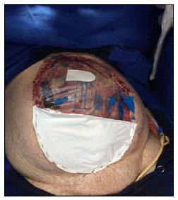
Initially described in 1984, plastic bags utilized to contain parenteral solutions were used to coat the abdominal opening on the patient for a second surgical intervention[12]. Afterwards, the polyvinyl chloride (plastic bag) was adopted to maintain the abdomen open, technique initially called as Bogota bag or Borraez’s bag[12]. It consists of a sterilized (by gas) plastic bag used for closure of abdominal wounds, being sutured to the skin or fascia of the anterior abdominal wall[13] (Figure 1). Variations of this treatment have already been described, and one of them is the use of a big bag, inside the peritoneal cavity, loose and that covers all of the abdominal viscera. Afterwards, the bag is placed under the skin and another plastic bag is inserted. That avoids many handle collisions from inside the abdominal cavity, adhesions between the viscera and parietal peritoneum, diminishing the risk of lesions and enabling cavity washing and its closure[12].
In general, it is very used in association with the polypropylene screen (as a reinforcement and contention), that is sutured to the skin, together with the edge of the collecting bag. This is done due to evisceration and difficulty in mobilizing the patient, consisting in the arising issues with the treatment[1].
However, it is a procedure that will require a larger use of stents and wash-ups, presenting a certain risk of eviscerations and difficulty in mobilizing the patient[12]. In addition, it generates skin lacerations, adherence of the intestine to the abdominal wall, difficulty in reproaching the abdomen and the need to gas sterilize the bag before its use, harder control of third space with minimum loss and any leak from under the bag can wet the bed increasing the risk of pneumonia[13,14].
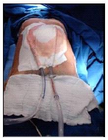
The technique developed by Barker et al[15] was described in 1995 using vacuum dressings for temporary closure of the abdominal cavity and since then it has been called “vacuum-pack” and became the most indicated for several conditions[16] (Figure 2). It was developed to be used with material found in the surgical rooms, presenting low cost and simple technique[15,17].
The vacuum-pack consists in an open polyethylene sheet between the abdominal viscera and anterior parietal peritoneum; a humid surgical dressing over the sheet with two suction drains. Then, an adhesive sheet throughout the wound, including a wide margin on the surrounding skin is placed. The stents are then connected to a suction device that enables 100-150 mmHg of continuum negative pressure[18]. This technique prevents damage to the abdominal wall due to the absence of sutures, preserving it to future approaches or definite closure, maintaining integrity of the fascia for later closure, allowing a quick approach to the abdominal cavity. The material utilized in contact with the abdominal viscera - the polyethylene sheet - is not adherent, and the vacuum-pack allows a better control of fluid quantity produced[17].
Regarding rates of fascial closure achieved with this technique, in 1997 a study obtained results of 61% of success. The patients, who were trauma victims and were subjected to Barker’s technique, had primary fascial closure on the 2nd laparotomy, being close to the achieved average[15]. Other studies had rates of 29%-100%[16,19-21]. The definite closure of the abdominal cavity after the 8th post-operative day from laparotomy was associated with a higher rate of complications[22].
Barker et al[23] showed their experience by using this technique in intestinal lesions submitted to resection in a study that lasted 11 years. There was no difference between patients who used this or other types of wound dressings regarding fistulas or leaks. Other studies reported fistula rates from 3% to 5%[19,23-25].
A study combining trauma victims and other cases of open abdomen, reports as existing complications: Abdominal abscess/infection, abdominal compartment syndrome, dehiscence, anastomotic leakage, coagulopathy, deep venous thrombosis, fascial necrosis, gastrointestinal ischemia, intestinal fistula, intestinal obstruction, pulmonary embolism and multiple organ failure[25]. In this study, it was not explained if these complications were directly related to the use of vacuum-pack. The complication rates related by Barker et al[15] are 15%.
The VAC, called VAC therapy or system is also known as negative pressure therapy (TPN), or sub atmospheric pressure (PTE) or as vacuum packing (TVV). Argenta and Morykwas[26] published an experimental work with VAC system, using acute wound in pigs, in 1997. In this work, they postulated that this system has a multimodal action mechanism. Since then, the clinical results observed in a variety of wounds were the best seen in comparison with the usual methods. The VAC therapy worked as a catalyst for the development of a series of studies[24,26]. Its efficiency in severe traumatic wounds helped to develop an area that belonged to plastic and reconstructive surgery[24].
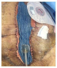
To apply this type of therapy, equipment made of reticulated foam is used and placed at the region and sealed in with the use of a patch (Figure 3). This foam can be made of polyurethane or polyvinyl alcohol, which generally presents 400-600 μm pores; it is cut to adjust perfectly along the wound before getting applied. If needed, several pieces of foam can be used to cover the gaps of the wound[24,27]. The foam is patched in with a drape that remains stuck on the skin with a 3 to 5 cm margin from the wound. In this sticker, a small 2 cm diameter hole is made in the center and a TRAC sticker (device that leads secretion to the tank) is linked to it. Then, a pump is connected to the foam (vacuum) that generates continuous sub atmospheric pressure (VAC-KCI, San Antonio, Texas, United States). Usually, putting an intermittent cycle of pressure for five minutes of suction and two minutes turned off. The pressure, in general, is adjusted to 125 mmHg and is distributed evenly throughout the wound through sponge pores. A plastic sticker is applied over the sponge to allow wound sealing[24,27-29].
Firstly, VAC removes stagnant fluid and debris and then is constantly optimizing blood supply and matrix deposition. Therefore, the partial oxygen pressure within the tissue increases and bacterial proliferation is reduced. Secondly, VAC leads to increased local interleukin-8 and vascular endothelial growth factor concentrations, which may trigger the accumulation of neutrophils and angiogenesis. With the cyclical application of sub atmospheric pressure, VAC alters the cytoskeleton of cells in the wound bed, triggering a cascade of intracellular signals that increase the rates of cell proliferation and division, and subsequent formation of granulation tissue[30].
The VAC system can be utilized for treatment of surgical infected wounds, trauma wounds, pressure ulcers, bone wounds and exposed hardware, diabetic foot ulcers and venous stasis ulcers. In addition, it can be used in reconstructing wounds, allowing elective planning of definite reconstructive surgery, without compromising the wound or the result. Furthermore, it increased significantly the success rate for skin graft when utilized as a cushion over the grafted wound[27].
Complications of VAC therapy are not frequent when the system is used correctly. Complication rates mostly described in literature are due to patient’s comorbidities and skin irritation caused by the drape. Other complications such as pain, infection or bleeding are not easily seen. Severe complications, such as toxic shock syndrome, anaerobic sepsis or thrombosis are described as well but rare[27].
The timing of primary conclusion with the vacuum assisted closure therapy is not always the fastest because it depends on each case, with the exception for patients with cardiovascular disease and/or diabetes. A portable VAC system (VAC freedom) is available in the market and it’s a regular size bag that has helped treating wounds carefully at home; this shows that VAC therapy improves quality of life because it reduces the length of hospital stay[31].
The total cost for VAC therapy is not significantly lower than the others; however, after analyzing time involvement and costs with nursing team, there is a considerable cut. Comfort for patients and nursing team are described in many cases as a relevant factor for choosing this therapy[31].
Comparing the use of VAC to other methods for temporary closure of the abdominal cavity, several studies showed that VAC performs better. A prospective study conducted by Batacchi et al[32] in 2009 compared patients who were abdominal trauma victims treated with the Bogota bag and VAC, during the abdominal cavity temporary closure. The VAC treatment was more effective in controlling intra-abdominal pressure (P < 0.01), normalization of serum lactate (P < 0.001), as well as less time needed for ventilation, faster abdominal closure and consequently shorter time at the intensive care unit and hospital stay. The results of the “Sequential Organ Failure Assessment” and mortality rate were not significantly different. Another study, conducted by Bee et al[33] compared polyglactin 910 (MESH) and VAC on temporary closure of the abdominal cavity after damage control surgery or compartment abdominal syndrome on a randomized study. In their results, VAC presented a 31% rate against 26% for the MESH group of late primary closure. The fistulas rates on the VAC group were 26% while on MESH, it was 5%. The differences presented in this study were not statistically different.
Kaplan et al[13] in 2005 concluded that VAC is the one to supply the ideal material for temporary closure on with greater satisfaction. From the 17 articles evaluated that had as object of study the 3 alternatives discussed in this article, the Bogota bag exhibited mortality rates of 53%, the vacuum-pack and VAC, 31% and 30%, respectively. Regarding complications such as fistulas, the VAC presented a rate of 2.6% against 7% of vacuum-pack and 13% of Bogota. Fascial closure was reached in 79% of patients subjected to VAC, while 58% was reached in vacuum-pack and Bogota bag reached only 18%[13].
Regarding intra-abdominal pressure (PIA), control Batacchi et al[32] in 2009, comparing the Bogota bag and VAC system, concluded that VAC was more effective on PIA control (P < 0.01) and on serum lactate (P < 0.001) during the first 24 h after surgical decompression[30]. The patients subjected to VAC had a faster abdominal closure and consequently stayed a shorter time at the intensive care unit; nevertheless, mortality rates were not different between the two groups.
Cheatham et al[25] in a 2013 study that compared VAC and vacuum-pack showed that both techniques present similar complication rates such as the development of abdominal compartment syndrome (8%) and fistulas (4%). VAC was associated with a bigger rate for primary facial closure longer than 30 d (73% against 27% of vacuum-pack) and smaller mortality rate on the same period for patients who needed open abdomen for at least 48 h. The difference in mortality between VAC and vacuum-pack increased significantly during the first 30 d due to the posterior development of multiple organ failure subjected to vacuum-pack and due to better peritoneal fluid removal rich in cytosines (increases organ dysfunction) through VAC[25]. Bruhin et al[34], in a recent study compared several techniques looking at contamination, fistula, and mortality rates, among others. Higher rates of primary fascial closure after VAC application in combination with “dynamic closure” technique (using traction, suture of dynamic retention or ABRA) were obtained. Moreover, patients who were not contaminated had a closure rate of 81%, followed by the exclusive use of VAC with rates of 72% and home-made (Barker technique) of 58%, taking into account that the Bogota bag data were inadequate[32]. Contaminated lesions were noticed, and resulted on higher rates of abdominal closure (74.6%), followed by its exclusive use (48%), Barker technique (35%) and Bogota bag (27%). In relation to the presence of fistulas and the mortality rate, the VAC technique had the lowest rate, being the highest value (40% of mortality rate) obtained by the Barker technique[32].
In conclusion, the treatment for open abdomen patients has evolved greatly in the last decades, with great improvement of the existing techniques and the emerging of new ones. Its main objective is to maximize tissue perfusion and to minimize potential intra-abdominal complications, such as fistulas and hernias, enabling early closure of the abdominal cavity. VAC therapy showed to be superior in relation to the other techniques, such as Bogota bag and vacuum-pack, allowing a better control of the third space fluid, avoiding complications such as fistula; it has lower mortality and infection rates, and better capacity in primary closure of the abdominal cavity. On the other hand, the Bogota bag technique was the least efficient, even though it is still very much used due to its low cost, easy access to material that is generally already present in the surgical room.
Laparostomy comprises a surgical approach where the abdominal wall is not sutured by plans and left open, allowing a regular inspection and drainage of intracavitary content. Its usage is indicated in cases of abdominal trauma, damage control surgery, sepsis of abdominal origin, severe acute pancreatitis, the need for new laparotomy, hemodynamic instability, retroperitoneal hematomas and necrotizing fasciitis, allowing a continuous evaluation of intra-abdominal pressure in order to avoid the development of abdominal compartment syndrome.
The different techniques available for the temporary closure presents several benefits as well as limitations for its use and today there is a tendency to use the properties of negative pressure to ensure a faster recover of the patient increasing the rate definite closure of the abdominal wall.
There are mainly three basic approaches for the maintenance of an open abdomen are: The Bogota bag technique presents as advantages the availability to material access, lower cost, fast and easily executed, and not dependent on a great experience from the surgeon. For allowing a great visibility and entrance in the abdominal cavity, it promotes greater control of the septic spots and enables several revisions. Moreover, it allows abdominal decompression, does not cause inflammatory reactions or allergies, being used in several parts of the organism and in any kind of surgery, from traumas to tumor removal. However, it presents higher rates of eviscerations and skin lacerations, implying greater need for abdominal drainage and wash-ups, higher rates of intestinal adhesion to the abdominal wall, difficulty in controlling the third space and leaks. Thus, it requires sterilization of the bag by gas preventing its availability. The technique proposed by Barker presents positive factors, such as easy assembly at a low cost, since it uses hospital material already used for other functions. Since acquiring these materials is easy, the technique application becomes simple. The closure of the abdominal cavity is done with less stress, preventing complications associated with moderate control of the fluid produced by the body. One disadvantage is the lack of efficiency in approaching the abdominal wall when the injury is extensive and important. There is a deficiency on maintaining pressure, which must be continuous and properly maintained in order to make the organs act in the recovery. In complicated cases with abdominal bleeding, it may take a while to detect this bleeding while draining the fluid. The temporary closure technique by negative pressure, the vacuum-assisted closure therapy (VAC), among all of them has a more effective action on closing the abdominal cavity because it reduces the tension on the injury boarders and works at cellular level increasing cell proliferation and division. Furthermore, it induces the formation of granulated tissue, reducing the aspect of matrix metalloproteinase on chronic wounds. There is an increase of blood flow at the site of the injury. At the same time protects the wound from external injuries to progress and removes stagnant fluids and debris, avoiding cavity contamination, reducing the interstitial pressure and bacterial proliferation. Thus, it promotes healing of the wound and decreases complication rates and hospital stay providing a better quality of life.
The treatment for open abdomen patients has evolved greatly in the last decades, with great improvement of the existing techniques and the emerging of new ones. Its main objective is to maximize tissue perfusion and to minimize potential intra-abdominal complications, such as fistulas and hernias, enabling early closure of the abdominal cavity. VAC therapy showed to be superior in relation to the other techniques, such as Bogota bag and vacuum-pack, allowing a better control of the third space fluid, avoiding complications such as fistula; it has lower mortality and infection rates, and better capacity in primary closure of the abdominal cavity.
Laparostomy comprises a surgical approach where the abdominal wall is not sutured by plans and left open, allowing a regular inspection and drainage of intracavitary content. Abdominal compartment syndrome is defined as a condition where organ dysfunction is caused by sustained intra-abdominal hypertension above 20 mmHg. However, it is known that values below this (> 12 mmHg) will also cause organ dysfunction, but to a lesser degree. The Bogota bag technique presents as advantages the availability to material access, lower cost, fast and easily executed, and not dependent on a great experience from the surgeon. For allowing a great visibility and entrance in the abdominal cavity, it promotes greater control of the septic spots and enables several revisions. The Barker’s technique presents positive factors, such as easy assembly at a low cost, since it uses hospital material already used for other functions such as surgical towels, sterile plastic bag and surgical drapes commercially available connected to the wall vacuum. The closure of the abdominal cavity is done with less stress, preventing complications associated with moderate control of the fluid produced by the body. The temporary closure technique by negative pressure, the VAC, reduces the tension on the injury boarders and works at cellular level increasing cell proliferation and division. Furthermore, it induces the formation of granulated tissue, reducing the aspect of matrix metalloproteinase on chronic wounds. At the same time protects the wound from external injuries to progress and removes stagnant fluids and debris using a continuous negative pressure applied by the system, avoiding cavity contamination, reducing the interstitial pressure and bacterial proliferation.
This article is a review about three different techniques of laparostomy, a technique that is nowadays very often utilized above all during damage control surgery. The subject of the review is quite interesting for emergency and trauma surgeons.
Manuscript source: Invited manuscript
Specialty type: Gastroenterology and hepatology
Country of origin: Brazil
Peer-review report classification
Grade A (Excellent): A
Grade B (Very good): 0
Grade C (Good): C
Grade D (Fair): 0
Grade E (Poor): 0
P- Reviewer: Aseni P, Pasalar M S- Editor: Gong XM L- Editor: A E- Editor: Wu HL
| 1. | Rotondo MF, Zonies DH. The damage control sequence and underlying logic. Surg Clin North Am. 1997;77:761-777. [RCA] [PubMed] [DOI] [Full Text] [Cited in This Article: ] [Cited by in Crossref: 322] [Cited by in RCA: 261] [Article Influence: 9.3] [Reference Citation Analysis (0)] |
| 2. | Coccolini F, Biffl W, Catena F, Ceresoli M, Chiara O, Cimbanassi S, Fattori L, Leppaniemi A, Manfredi R, Montori G. The open abdomen, indications, management and definitive closure. World J Emerg Surg. 2015;10:32. [RCA] [PubMed] [DOI] [Full Text] [Full Text (PDF)] [Cited in This Article: ] [Cited by in Crossref: 82] [Cited by in RCA: 77] [Article Influence: 7.7] [Reference Citation Analysis (0)] |
| 3. | Starling SV, Reis MCW. Utilização do Sistema VAC em Peritoniostomia. Intergastro e Trauma. 2013;24:271-277. [Cited in This Article: ] |
| 4. | Rasilainen SK, Mentula PJ, Leppäniemi AK. Vacuum and mesh-mediated fascial traction for primary closure of the open abdomen in critically ill surgical patients. Br J Surg. 2012;99:1725-1732. [RCA] [PubMed] [DOI] [Full Text] [Cited in This Article: ] [Cited by in Crossref: 104] [Cited by in RCA: 111] [Article Influence: 8.5] [Reference Citation Analysis (0)] |
| 5. | Zeni M, G Junior RL, Silva AB. Síndrome compartimental abdominal: Rotinas do serviço de cirurgia geral do Hospital Governador Celso Ramos. Acm Arq catarin med. 2010;39. [Cited in This Article: ] |
| 6. | Ferreira F, Barbosa E, Guerreiro E, Fraga GP, Nascimento Junior B, Rizoli S. Fechamento sequencial da parede abdominal com tração fascial contínua (mediada por tela ou sutura) e terapia a vácuo. Rev Col Bras Cir. 2013;40:85-89. [Cited in This Article: ] |
| 7. | Maia DEF, Ribeiro Junior MAF. Manual de condutas básicas em cirurgia. Controle de danos. 1st ed. Santos: Gen Roca 2013; 126-129. [Cited in This Article: ] |
| 8. | Refinetti RA, Martinez R. Pancreatite Necro-hemorrágica: Atualização e momento de operar. Abcd Arq Bras Cir Dig. 2010;23:122-127. [RCA] [DOI] [Full Text] [Cited in This Article: ] [Cited by in Crossref: 2] [Cited by in RCA: 2] [Article Influence: 0.1] [Reference Citation Analysis (0)] |
| 9. | Utiyama EM, Pflug ARM, Damous SHB, Rodrigues Junior AC, Montero EFS, Birolini CAV. Fechamento abdominal temporário com dispositivo tela-zíper para tratamento da sepse intra-abdominal. Rev Col Bras Cir. 2015;42:18-24. [RCA] [DOI] [Full Text] [Cited in This Article: ] [Cited by in Crossref: 8] [Cited by in RCA: 8] [Article Influence: 0.8] [Reference Citation Analysis (0)] |
| 10. | Edelmuth RCL, Buscariolli YS, Ribeiro Junior MAF. Cirurgia para controle de danos: Estado atual. Rev Col Bras Cir. 2013;40:142-151. [Cited in This Article: ] |
| 11. | Borraez AO. Abdomen abierto: la herida más desafiante. Rev Colomb Cir. 2008;23:204-209. [Cited in This Article: ] |
| 12. | Torres NJR, Barreto AP, Prudente ACL, Santos AM, Santiago RR. Uso da peritoneostomia na sepse abdominal. Rev Bras Coloproctol. 2007;27:278-283. [Cited in This Article: ] |
| 13. | Kaplan M, Banwell P, Orgill DP, Ivatury RR, Demetriades VD, Moore FA, Miller P, Nicholes J, Henry S. Guidelines for the Management of the Open Abdomen. Wounds. 2005;17 Suppl S1:1-27. [Cited in This Article: ] |
| 14. | Ferraz ED, Vieira OM. Técnica de fechamento progressivo na laparostomia e descompressão abdominal. Rev Col Bras Cir. 2000;27:237-244. [RCA] [DOI] [Full Text] [Cited in This Article: ] [Cited by in Crossref: 3] [Cited by in RCA: 3] [Article Influence: 0.1] [Reference Citation Analysis (0)] |
| 15. | Barker DE, Green JM, Maxwell RA, Smith PW, Mejia VA, Dart BW, Cofer JB, Roe SM, Burns RP. Experience with vacuum-pack temporary abdominal wound closure in 258 trauma and general and vascular surgical patients. J Am Coll Surg. 2007;204:784-792; discussion 792-793. [RCA] [PubMed] [DOI] [Full Text] [Cited in This Article: ] [Cited by in Crossref: 143] [Cited by in RCA: 134] [Article Influence: 7.4] [Reference Citation Analysis (0)] |
| 16. | Kreis BE, de Mol van Otterloo AJ, Kreis RW. Open abdomen management: a review of its history and a proposed management algorithm. Med Sci Monit. 2013;19:524-533. [RCA] [PubMed] [DOI] [Full Text] [Full Text (PDF)] [Cited in This Article: ] [Cited by in Crossref: 56] [Cited by in RCA: 58] [Article Influence: 4.8] [Reference Citation Analysis (0)] |
| 17. | Rezende-Neto JB, Cunha-Melo JR, Andrade MV. Vacuum pack technique for temporary abdominal wound closure. Rev Col Bras Cir. 2007;34:336-339. [Cited in This Article: ] |
| 18. | Smith LA, Barker DE, Chase CW, Somberg LB, Brock WB, Burns RP. Vacuum pack technique of temporary abdominal closure: a four-year experience. Am Surg. 1997;63:1102-1107; discussion 1107-1108. [PubMed] [Cited in This Article: ] |
| 19. | Navsaria PH, Bunting M, Omoshoro-Jones J, Nicol AJ, Kahn D. Temporary closure of open abdominal wounds by the modified sandwich-vacuum pack technique. Br J Surg. 2003;90:718-722. [RCA] [PubMed] [DOI] [Full Text] [Cited in This Article: ] [Cited by in Crossref: 100] [Cited by in RCA: 96] [Article Influence: 4.4] [Reference Citation Analysis (0)] |
| 20. | Boele van Hensbroek P, Wind J, Dijkgraaf MG, Busch OR, Goslings JC. Temporary closure of the open abdomen: a systematic review on delayed primary fascial closure in patients with an open abdomen. World J Surg. 2009;33:199-207. [RCA] [PubMed] [DOI] [Full Text] [Full Text (PDF)] [Cited in This Article: ] [Cited by in Crossref: 214] [Cited by in RCA: 192] [Article Influence: 12.0] [Reference Citation Analysis (0)] |
| 21. | Cothren CC, Moore EE, Johnson JL, Moore JB, Burch JM. One hundred percent fascial approximation with sequential abdominal closure of the open abdomen. Am J Surg. 2006;192:238-242. [RCA] [PubMed] [DOI] [Full Text] [Cited in This Article: ] [Cited by in Crossref: 132] [Cited by in RCA: 120] [Article Influence: 6.3] [Reference Citation Analysis (0)] |
| 22. | Miller RS, Morris JA, Diaz JJ, Herring MB, May AK. Complications after 344 damage-control open celiotomies. J Trauma. 2005;59:1365-1371; discussion 1371-1374. [RCA] [PubMed] [DOI] [Full Text] [Cited in This Article: ] [Cited by in Crossref: 229] [Cited by in RCA: 195] [Article Influence: 10.3] [Reference Citation Analysis (0)] |
| 23. | Barker DE, Kaufman HJ, Smith LA, Ciraulo DL, Richart CL, Burns RP. Vacuum pack technique of temporary abdominal closure: a 7-year experience with 112 patients. J Trauma. 2000;48:201-206; discussion 206-207. [RCA] [PubMed] [DOI] [Full Text] [Cited in This Article: ] [Cited by in Crossref: 332] [Cited by in RCA: 273] [Article Influence: 10.9] [Reference Citation Analysis (0)] |
| 24. | Banwell PE, Téot L. Topical negative pressure (TNP): the evolution of a novel wound therapy. J Wound Care. 2003;12:22-28. [RCA] [PubMed] [DOI] [Full Text] [Cited in This Article: ] [Cited by in Crossref: 47] [Cited by in RCA: 45] [Article Influence: 2.3] [Reference Citation Analysis (0)] |
| 25. | Cheatham ML, Demetriades D, Fabian TC, Kaplan MJ, Miles WS, Schreiber MA, Holcomb JB, Bochicchio G, Sarani B, Rotondo MF. Prospective study examining clinical outcomes associated with a negative pressure wound therapy system and Barker’s vacuum packing technique. World J Surg. 2013;37:2018-2030. [RCA] [PubMed] [DOI] [Full Text] [Full Text (PDF)] [Cited in This Article: ] [Cited by in Crossref: 118] [Cited by in RCA: 113] [Article Influence: 10.3] [Reference Citation Analysis (0)] |
| 26. | Argenta LC, Morykwas MJ. Vacuum-assisted closure: a new method for wound control and treatment: clinical experience. Ann Plast Surg. 1997;38:563-576; discussion 577. [RCA] [PubMed] [DOI] [Full Text] [Cited in This Article: ] [Cited by in Crossref: 1531] [Cited by in RCA: 1358] [Article Influence: 48.5] [Reference Citation Analysis (0)] |
| 27. | Leijnen M, Steenvoorde P, Vandoorn I, Da costa SA, Oskam J. A portable vacuum device improves upon this 20-year-old therapy. J Wound Care. 2007;16:211-212. [RCA] [DOI] [Full Text] [Cited in This Article: ] [Cited by in Crossref: 6] [Cited by in RCA: 7] [Article Influence: 0.4] [Reference Citation Analysis (0)] |
| 28. | Labler L, Keel M, Trentz O, Heinzelmann M. Wound conditioning by vacuum assisted closure (V.A.C.) in postoperative infections after dorsal spine surgery. Eur Spine J. 2006;15:1388-1396. [RCA] [PubMed] [DOI] [Full Text] [Cited in This Article: ] [Cited by in Crossref: 44] [Cited by in RCA: 52] [Article Influence: 2.7] [Reference Citation Analysis (0)] |
| 29. | Milcheski DA, Zampieri FMC, Nakamoto HA, Tuma Jr P, Ferreira MC. Terapia por pressão negativa na ferida traumática complexa do períneo. Rev Col Bras Cir. 2013;40:312-317. [Cited in This Article: ] |
| 30. | Wang W, Pan Z, Hu X, Li Z, Zhao Y, Yu AX. Vacuum-assisted closure increases ICAM-1, MIF, VEGF and collagen I expression in wound therapy. Exp Ther Med. 2014;7:1221-1226. [RCA] [PubMed] [DOI] [Full Text] [Full Text (PDF)] [Cited in This Article: ] [Cited by in Crossref: 13] [Cited by in RCA: 15] [Article Influence: 1.4] [Reference Citation Analysis (0)] |
| 31. | Braakenburg A, Obdeijn MC, Feitz R, van Rooij IA, van Griethuysen AJ, Klinkenbijl JH. The clinical efficacy and cost effectiveness of the vacuum-assisted closure technique in the management of acute and chronic wounds: a randomized controlled trial. Plast Reconstr Surg. 2006;118:390-397; discussion 398-400. [RCA] [PubMed] [DOI] [Full Text] [Cited in This Article: ] [Cited by in Crossref: 216] [Cited by in RCA: 208] [Article Influence: 10.9] [Reference Citation Analysis (0)] |
| 32. | Batacchi S, Matano S, Nella A, Zagli G, Bonizzoli M, Pasquini A, Anichini V, Tucci V, Manca G, Ban K. Vacuum-assisted closure device enhances recovery of critically ill patients following emergency surgical procedures. Crit Care. 2009;13:R194. [RCA] [PubMed] [DOI] [Full Text] [Full Text (PDF)] [Cited in This Article: ] [Cited by in Crossref: 54] [Cited by in RCA: 59] [Article Influence: 3.7] [Reference Citation Analysis (0)] |
| 33. | Bee TK, Croce MA, Magnotti LJ, Zarzaur BL, Maish GO, Minard G, Schroeppel TJ, Fabian TC. Temporary abdominal closure techniques: a prospective randomized trial comparing polyglactin 910 mesh and vacuum-assisted closure. J Trauma. 2008;65:337-342; discussion 342-344. [RCA] [PubMed] [DOI] [Full Text] [Cited in This Article: ] [Cited by in Crossref: 122] [Cited by in RCA: 140] [Article Influence: 8.2] [Reference Citation Analysis (0)] |
| 34. | Bruhin A, Ferreira F, Chariker M, Smith J, Runkel N. Systematic review and evidence based recommendations for the use of negative pressure wound therapy in the open abdomen. Int J Surg. 2014;12:1105-1114. [RCA] [PubMed] [DOI] [Full Text] [Cited in This Article: ] [Cited by in Crossref: 95] [Cited by in RCA: 90] [Article Influence: 8.2] [Reference Citation Analysis (0)] |


