Copyright
©The Author(s) 2016.
World J Gastrointest Surg. Jun 27, 2016; 8(6): 407-423
Published online Jun 27, 2016. doi: 10.4240/wjgs.v8.i6.407
Published online Jun 27, 2016. doi: 10.4240/wjgs.v8.i6.407
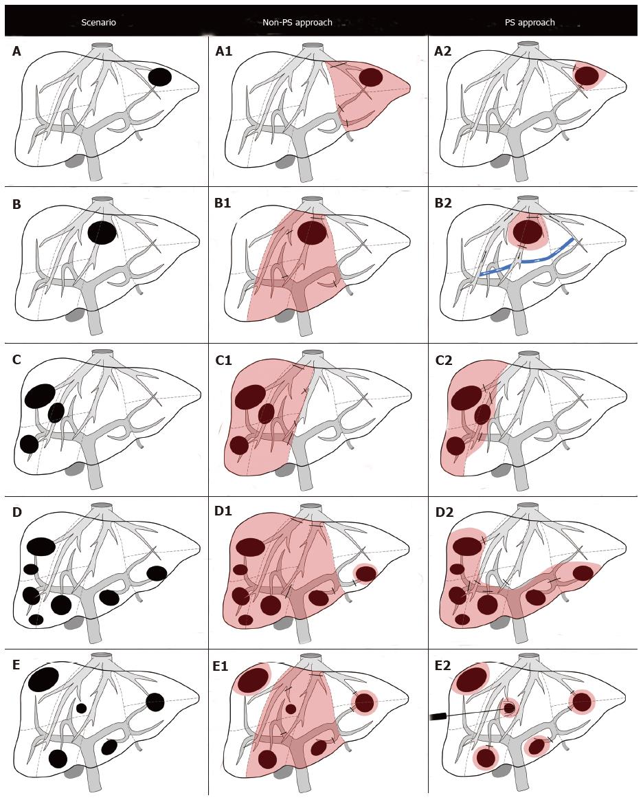
Figure 3 Diagram of non-segment-oriented parenchymal-sparing resections according to different surgical scenarios.
A: Metastatic lesion in S2; A1: Left lateral sectionectomy; A2: Atypical resection of S2; B: Metastatic lesion infiltrating the MHV close to the hepato-caval confluence; B1: Central hepatectomy; B2: Mini-mesohepatectomy is possible due to the presence of communicating hepatic veins; C: Metastatic lesions in right posterior section invading the RHV and tumor in proximity of right anterior portal branch; C1: Right hepatectomy; C2: Systematic extended right posterior sectionectomy; D: Liver metastases peripherally located in S3, 4b, 5, 6 and 7; D1: Right trisectionectomy; D2: Lower inferior hepatectomy; E: Multiple bilateral metastases; E1: Atypical resections combined with central hepatectomy; E2: Multiple atypical resections combined with radiofrequency ablation. PS: Parenchymal-sparing; RHV: Right hepatic vein; MHV: Middle hepatic vein.
- Citation: Alvarez FA, Sanchez Claria R, Oggero S, de Santibañes E. Parenchymal-sparing liver surgery in patients with colorectal carcinoma liver metastases. World J Gastrointest Surg 2016; 8(6): 407-423
- URL: https://www.wjgnet.com/1948-9366/full/v8/i6/407.htm
- DOI: https://dx.doi.org/10.4240/wjgs.v8.i6.407


