Copyright
©The Author(s) 2016.
World J Gastrointest Surg. Nov 27, 2016; 8(11): 729-734
Published online Nov 27, 2016. doi: 10.4240/wjgs.v8.i11.729
Published online Nov 27, 2016. doi: 10.4240/wjgs.v8.i11.729
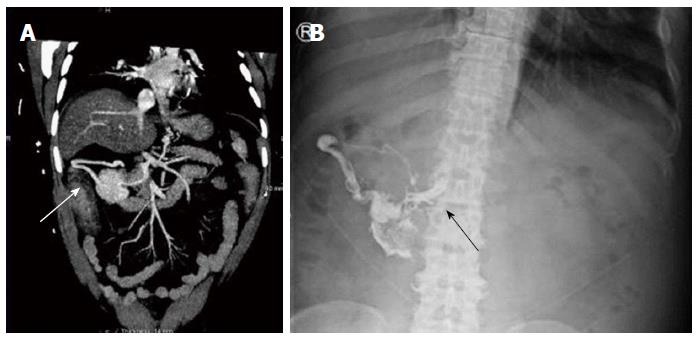
Figure 2 Duodenal varix on computed tomography and plain radiograph before and after glue injection.
A: Computed tomography angiogram showing a large abdominal varix meeting the duodenum (white arrow); B: After glue injection a plain radiograph showed lipiodol/glue in the same vessel, with extension medially up to the portal vein (black arrow).
- Citation: Al-Hillawi L, Wong T, Tritto G, Berry PA. Pitfalls in histoacryl glue injection therapy for oesophageal, gastric and ectopic varices: A review. World J Gastrointest Surg 2016; 8(11): 729-734
- URL: https://www.wjgnet.com/1948-9366/full/v8/i11/729.htm
- DOI: https://dx.doi.org/10.4240/wjgs.v8.i11.729


