Copyright
©The Author(s) 2015.
World J Gastrointest Surg. Jul 27, 2015; 7(7): 116-122
Published online Jul 27, 2015. doi: 10.4240/wjgs.v7.i7.116
Published online Jul 27, 2015. doi: 10.4240/wjgs.v7.i7.116
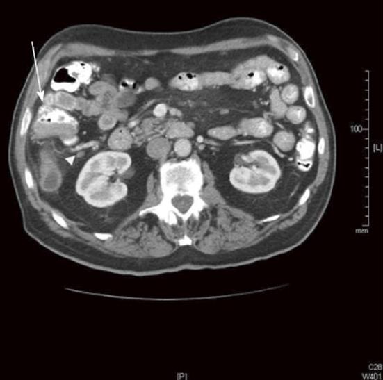
Figure 2 Computed tomography image showing positive nodal disease.
This computed tomography image read by outside radiologist as lymph node (LN) negative disease was confirmed to be LN positive by final pathology (arrow head). Contiguous with the base of the appendix, an irregular cecal soft tissue mass (4.5 cm × 2.2 cm × 3.2 cm) can be seen (arrow).
- Citation: Choi AH, Nelson RA, Schoellhammer HF, Cho W, Ko M, Arrington A, Oxner CR, Fakih M, Wong J, Sentovich SM, Garcia-Aguilar J, Kim J. Accuracy of computed tomography in nodal staging of colon cancer patients. World J Gastrointest Surg 2015; 7(7): 116-122
- URL: https://www.wjgnet.com/1948-9366/full/v7/i7/116.htm
- DOI: https://dx.doi.org/10.4240/wjgs.v7.i7.116


