Copyright
©2014 Baishideng Publishing Group Inc.
World J Gastrointest Surg. May 27, 2014; 6(5): 84-87
Published online May 27, 2014. doi: 10.4240/wjgs.v6.i5.84
Published online May 27, 2014. doi: 10.4240/wjgs.v6.i5.84
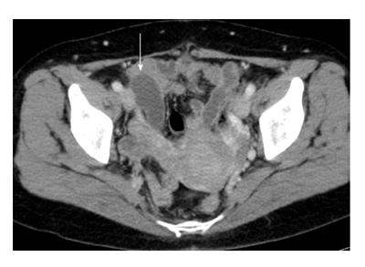
Figure 1 Computed tomography.
The arrow shows a 55 mm × 25 mm, low-density and no-contrast lesion at the appendix.
- Citation: Fujino S, Miyoshi N, Noura S, Shingai T, Tomita Y, Ohue M, Yano M. Single-incision laparoscopic cecectomy for low-grade appendiceal mucinous neoplasm after laparoscopic rectectomy. World J Gastrointest Surg 2014; 6(5): 84-87
- URL: https://www.wjgnet.com/1948-9366/full/v6/i5/84.htm
- DOI: https://dx.doi.org/10.4240/wjgs.v6.i5.84


