Copyright
©2014 Baishideng Publishing Group Co.
World J Gastrointest Surg. Apr 27, 2014; 6(4): 70-73
Published online Apr 27, 2014. doi: 10.4240/wjgs.v6.i4.70
Published online Apr 27, 2014. doi: 10.4240/wjgs.v6.i4.70
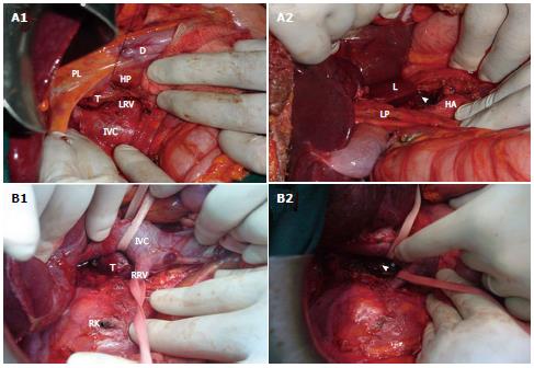
Figure 2 Operative view.
A: Patient 1. A1: Separation of the tumor (T) from the anterior aspect of the inferior vena cava (IVC); A2: Surgical site after tumor resection (arrowhead); B: Patient 3. B1: Separation of the tumor (T) from the posterior wall of the IVC; B2: Surgical site after tumor resection (arrowhead). LRV: Left renal vein; D: Duodenum; HP: Head of the pancreas; PL: Liver pedicle; RRV: Right renal vein; RK: Right kidney; HA: Hepatic artery; LP: Liver pedicle; L: Liver (Lobe of Spiegel).
- Citation: Kallel H, Hentati H, Baklouti A, Gassara A, Saadaoui A, Halek G, Landolsi S, Ouaer ME, Chaieb W, Maamouri F, Mannaï S. Retroperitoneal paragangliomas: Report of 4 cases. World J Gastrointest Surg 2014; 6(4): 70-73
- URL: https://www.wjgnet.com/1948-9366/full/v6/i4/70.htm
- DOI: https://dx.doi.org/10.4240/wjgs.v6.i4.70


