Copyright
©2014 Baishideng Publishing Group Co.
World J Gastrointest Surg. Apr 27, 2014; 6(4): 70-73
Published online Apr 27, 2014. doi: 10.4240/wjgs.v6.i4.70
Published online Apr 27, 2014. doi: 10.4240/wjgs.v6.i4.70
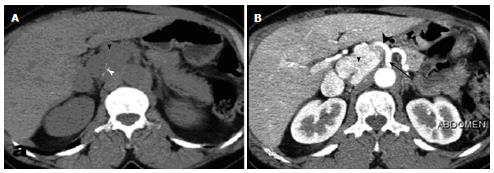
Figure 1 Abdominal computed tomography without (A) and after (B) contrast material administration, showing the tumor (arrowhead) with calcifications (white arrowhead) and precocious enhancement.
Note the close tumoral relationship to the celiac trunk (black arrow) and hepatic artery (black arrowhead).
- Citation: Kallel H, Hentati H, Baklouti A, Gassara A, Saadaoui A, Halek G, Landolsi S, Ouaer ME, Chaieb W, Maamouri F, Mannaï S. Retroperitoneal paragangliomas: Report of 4 cases. World J Gastrointest Surg 2014; 6(4): 70-73
- URL: https://www.wjgnet.com/1948-9366/full/v6/i4/70.htm
- DOI: https://dx.doi.org/10.4240/wjgs.v6.i4.70


