Copyright
©2014 Baishideng Publishing Group Co.
World J Gastrointest Surg. Apr 27, 2014; 6(4): 65-69
Published online Apr 27, 2014. doi: 10.4240/wjgs.v6.i4.65
Published online Apr 27, 2014. doi: 10.4240/wjgs.v6.i4.65
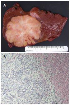
Figure 2 Photograph.
A: Right lobe liver resection specimen showing a 6.0 cm × 5.5 cm × 5 cm well-circumscribed tumor with a firm, heterogeneous, yellow tan and white cut surface with areas of fibrosis. The surrounding liver is unremarkable; B: Representative photomicrograph of the tumor shows anastomosing glandular structures composed of highly pleomorphic epithelial cells in a desmoplastic stroma. The findings are consistent with intrahepatic cholangiocarcinoma (HE, original magnification × 200).
- Citation: Labgaa I, Carrasco-Avino G, Fiel MI, Schwartz ME. Pancreatic recurrence of intrahepatic cholangiocarcinoma: Case report and review of the literature. World J Gastrointest Surg 2014; 6(4): 65-69
- URL: https://www.wjgnet.com/1948-9366/full/v6/i4/65.htm
- DOI: https://dx.doi.org/10.4240/wjgs.v6.i4.65


