Copyright
©2014 Baishideng Publishing Group Co.
World J Gastrointest Surg. Mar 27, 2014; 6(3): 42-46
Published online Mar 27, 2014. doi: 10.4240/wjgs.v6.i3.42
Published online Mar 27, 2014. doi: 10.4240/wjgs.v6.i3.42
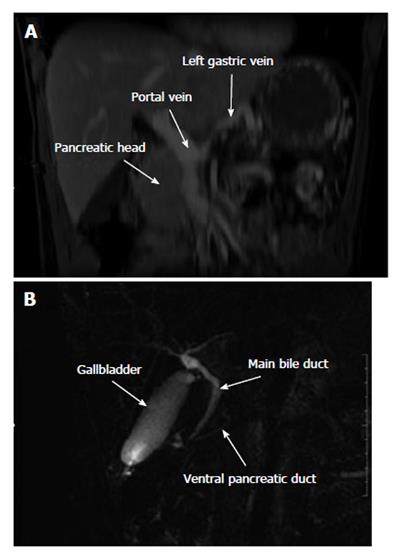
Figure 4 Coronal magnetic resonance imagery showing lack of the body and tail of the pancreas and of the splenic vein (A), magnetic resonance cholangiopancreatography showing the common bile duct joining the ventral pancreatic duct at the posterior part of the head of the pancreas (B).
The dorsal pancreatic duct is not visible.
- Citation: Gagnière J, Dupré A, Ines DD, Tixier L, Pezet D, Buc E. Giant mucinous cystic adenoma with pancreatic atrophy mimicking dorsal agenesis of the pancreas. World J Gastrointest Surg 2014; 6(3): 42-46
- URL: https://www.wjgnet.com/1948-9366/full/v6/i3/42.htm
- DOI: https://dx.doi.org/10.4240/wjgs.v6.i3.42


