Copyright
©2014 Baishideng Publishing Group Co.
World J Gastrointest Surg. Mar 27, 2014; 6(3): 42-46
Published online Mar 27, 2014. doi: 10.4240/wjgs.v6.i3.42
Published online Mar 27, 2014. doi: 10.4240/wjgs.v6.i3.42
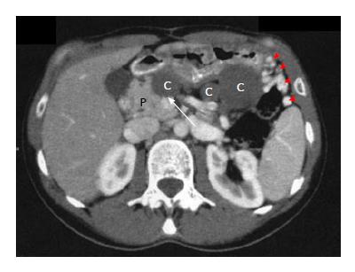
Figure 3 Post-operative contrast-enhanced computed tomography showing three low-density homogenous cystic lesions suggesting secondary dissemination of the resected cyst.
Major tributaries were also present around the stomach (red arrowheads). The P has a total distal atrophy as shown by lack of pancreatic tissue behind the mesenterico-portal axis (white arrow). P: Pancreas; C: Cyst.
- Citation: Gagnière J, Dupré A, Ines DD, Tixier L, Pezet D, Buc E. Giant mucinous cystic adenoma with pancreatic atrophy mimicking dorsal agenesis of the pancreas. World J Gastrointest Surg 2014; 6(3): 42-46
- URL: https://www.wjgnet.com/1948-9366/full/v6/i3/42.htm
- DOI: https://dx.doi.org/10.4240/wjgs.v6.i3.42


