Copyright
©2014 Baishideng Publishing Group Inc.
World J Gastrointest Surg. Dec 27, 2014; 6(12): 253-258
Published online Dec 27, 2014. doi: 10.4240/wjgs.v6.i12.253
Published online Dec 27, 2014. doi: 10.4240/wjgs.v6.i12.253
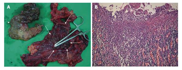
Figure 3 Gross anatomic specimen and its macroscopic histology show severe tuberculous inflammation and necrosis from the totally resected stomach and a part of duodenum.
A: A huge defect (white arrows) in the gastric wall along the midbody and fundus. The dirty ragged tissue (white arrowhead) which was covering the defect was identified to be the infracted gastric wall pathologically; B: Histologic findings of the resected specimen in hematoxylin and eosin staining (original magnification × 100) showing diffuse transmural inflammation with extensive multiple ulcers on the stomach.
- Citation: Park CS, Seo KW, Park CR, Nah YW, Suh JH. Case of bronchoesophageal fistula with gastric perforation due to multidrug-resistant tuberculosis. World J Gastrointest Surg 2014; 6(12): 253-258
- URL: https://www.wjgnet.com/1948-9366/full/v6/i12/253.htm
- DOI: https://dx.doi.org/10.4240/wjgs.v6.i12.253


