Copyright
©2014 Baishideng Publishing Group Inc.
World J Gastrointest Surg. Dec 27, 2014; 6(12): 253-258
Published online Dec 27, 2014. doi: 10.4240/wjgs.v6.i12.253
Published online Dec 27, 2014. doi: 10.4240/wjgs.v6.i12.253
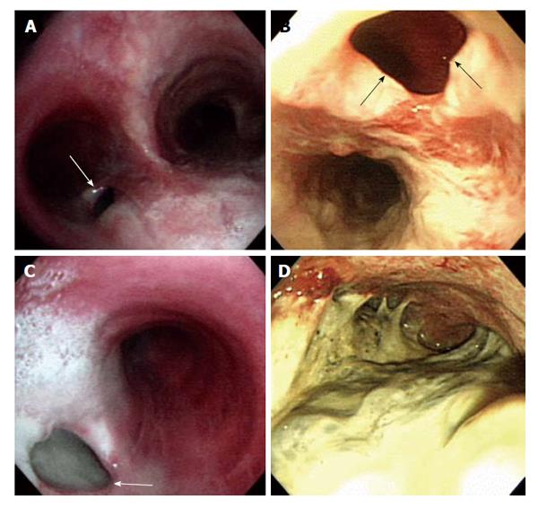
Figure 2 Bronchoscopy (A and C) and esophagogastroduodenoscopy (B and D) show tuberculous bronchoesophageal fistula and nearby lesions.
A: A 1.0 cm × 1.5 cm sized hole (white arrow) was seen at the inferior wall aspect of the left main bronchus; B: Esophagogastroduodenoscopy showing a large fistula (black arrows) was noted at the level of incisor 28 cm below; C: The lumen of fistula (white arrow) was filled up with whitish exudate secretion and materials; D: Edematous and hyperemic inflammatory mucosal changes with exudate materials were observed at whole stomach.
- Citation: Park CS, Seo KW, Park CR, Nah YW, Suh JH. Case of bronchoesophageal fistula with gastric perforation due to multidrug-resistant tuberculosis. World J Gastrointest Surg 2014; 6(12): 253-258
- URL: https://www.wjgnet.com/1948-9366/full/v6/i12/253.htm
- DOI: https://dx.doi.org/10.4240/wjgs.v6.i12.253


