Copyright
©2014 Baishideng Publishing Group Inc.
World J Gastrointest Surg. Dec 27, 2014; 6(12): 248-252
Published online Dec 27, 2014. doi: 10.4240/wjgs.v6.i12.248
Published online Dec 27, 2014. doi: 10.4240/wjgs.v6.i12.248
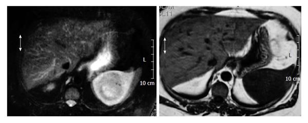
Figure 2 Axial T2W fat sat image shows a large intrasplenic mass.
Notice the slightly decrease of signal intensity and the lack of a peripherical capsule.
- Citation: Prieto-Nieto MI, Pérez-Robledo JP, Díaz-San Andrés B, Nistal M, Rodríguez-Montes JA. Inflammatory pseudotumour of the spleen associated with splenic tuberculosis. World J Gastrointest Surg 2014; 6(12): 248-252
- URL: https://www.wjgnet.com/1948-9366/full/v6/i12/248.htm
- DOI: https://dx.doi.org/10.4240/wjgs.v6.i12.248


