Copyright
©2013 Baishideng Publishing Group Co.
World J Gastrointest Surg. Sep 27, 2013; 5(9): 256-258
Published online Sep 27, 2013. doi: 10.4240/wjgs.v5.i9.256
Published online Sep 27, 2013. doi: 10.4240/wjgs.v5.i9.256
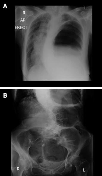
Figure 1 Plain radiographic images of the patient upon emergency admission.
A: Erect chest radiograph upon admission demonstrated that the patient had a collapsed left lung and elevated left hemidiaphragm with a large viscous identified under the left hemidiaphragm; B: Plain abdominal radiograph on admission revealed a gas filled viscous within the left upper quadrant consistent with caecal volvulus.
- Citation: Bhogal RH, Maleki K, Patel R. Colonic tumour precipitating caecal volvulus within a diaphragmatic hernia. World J Gastrointest Surg 2013; 5(9): 256-258
- URL: https://www.wjgnet.com/1948-9366/full/v5/i9/256.htm
- DOI: https://dx.doi.org/10.4240/wjgs.v5.i9.256


