Copyright
©2013 Baishideng Publishing Group Co.
World J Gastrointest Surg. Jul 27, 2013; 5(7): 229-232
Published online Jul 27, 2013. doi: 10.4240/wjgs.v5.i7.229
Published online Jul 27, 2013. doi: 10.4240/wjgs.v5.i7.229
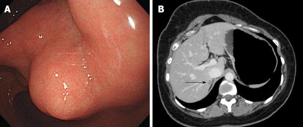
Figure 1 Esophagogastroduodenoscopy.
A: Gastroendoscopy revealed an antral submucosal tumor without ulceration and hemorrhage; B: Computed tomography revealed a hypoattenuating nodule in segment VII of the liver, with peripheral weak enhancement in the portal phase (arrow).
- Citation: Hong SW, Lee WY, Lee HK. Hepatic paraganglioma and multifocal gastrointestinal stromal tumor in a female: Incomplete Carney triad. World J Gastrointest Surg 2013; 5(7): 229-232
- URL: https://www.wjgnet.com/1948-9366/full/v5/i7/229.htm
- DOI: https://dx.doi.org/10.4240/wjgs.v5.i7.229


