Copyright
©2013 Baishideng Publishing Group Co.
World J Gastrointest Surg. Jun 27, 2013; 5(6): 195-198
Published online Jun 27, 2013. doi: 10.4240/wjgs.v5.i6.195
Published online Jun 27, 2013. doi: 10.4240/wjgs.v5.i6.195
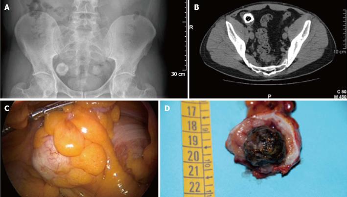
Figure 1 Abdominal pain.
A: Lower abdomen X-ray shows a rounded radio-opaque formation in the right iliac fossa; B: Lower abdomen computed tomography showing a roundish-filling defect in right iliac fossa; C: Laparoscopic intraoperative view of the dilated appendix due to the presence of a foreign body; D: The marrowbone included in a calcified fecaloma.
- Citation: Antonacci N, Labombarda M, Ricci C, Buscemi S, Casadei R, Minni F. A bizarre foreign body in the appendix: A case report. World J Gastrointest Surg 2013; 5(6): 195-198
- URL: https://www.wjgnet.com/1948-9366/full/v5/i6/195.htm
- DOI: https://dx.doi.org/10.4240/wjgs.v5.i6.195


