Copyright
©2013 Baishideng Publishing Group Co.
World J Gastrointest Surg. Mar 27, 2013; 5(3): 62-67
Published online Mar 27, 2013. doi: 10.4240/wjgs.v5.i3.62
Published online Mar 27, 2013. doi: 10.4240/wjgs.v5.i3.62
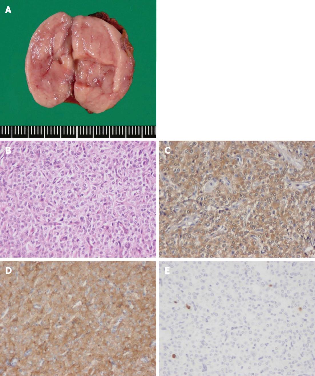
Figure 2 Macroscopic findings and pathological features of the resected tumor.
A: Gross findings of the resected specimen. The tumor was encapsulated and measured 3 cm × 1.5 cm × 1.5 cm; B: The paraganglioma comprised a dual cell population arranged in a characteristic nested Zellballen pattern (HE stain, × 400); C: Immunohistochemistory of Chromogranin A, × 400; D: Synaptophysin were strongly positive and confirmed a neuroendocrine origin, supporting the diagnosis of paraganglioma, × 400; E: The MIB-1 labeling index, × 400.
- Citation: Fujita T, Kamiya K, Takahashi Y, Miyazaki S, Iino I, Kikuchi H, Hiramatsu Y, Ohta M, Baba S, Konno H. Mesenteric paraganglioma: Report of a case. World J Gastrointest Surg 2013; 5(3): 62-67
- URL: https://www.wjgnet.com/1948-9366/full/v5/i3/62.htm
- DOI: https://dx.doi.org/10.4240/wjgs.v5.i3.62


