Copyright
©2013 Baishideng Publishing Group Co.
World J Gastrointest Surg. Oct 27, 2013; 5(10): 278-281
Published online Oct 27, 2013. doi: 10.4240/wjgs.v5.i10.278
Published online Oct 27, 2013. doi: 10.4240/wjgs.v5.i10.278
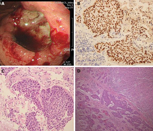
Figure 2 The biopsy showed syncytial growth of polygonal shaped poorly differentiated cells.
A: Macroscopically, there was an ulcerofungating 4.0 cm × 3.5 cm lesion on the body; B, C: The bronchoscopic biopsy showed growth of polygonal shaped cells which were strongly positive for p63 and consistent with squamous cell carcinoma; D: Gastric ulcer specimen revealed multiple lymphatic invasion of carcinoma and chronic gastritis.
- Citation: Kim YI, Kang BC, Sung SH. Surgically resected gastric metastasis of pulmonary squamous cell carcinoma. World J Gastrointest Surg 2013; 5(10): 278-281
- URL: https://www.wjgnet.com/1948-9366/full/v5/i10/278.htm
- DOI: https://dx.doi.org/10.4240/wjgs.v5.i10.278


