Copyright
©2013 Baishideng Publishing Group Co.
World J Gastrointest Surg. Oct 27, 2013; 5(10): 278-281
Published online Oct 27, 2013. doi: 10.4240/wjgs.v5.i10.278
Published online Oct 27, 2013. doi: 10.4240/wjgs.v5.i10.278
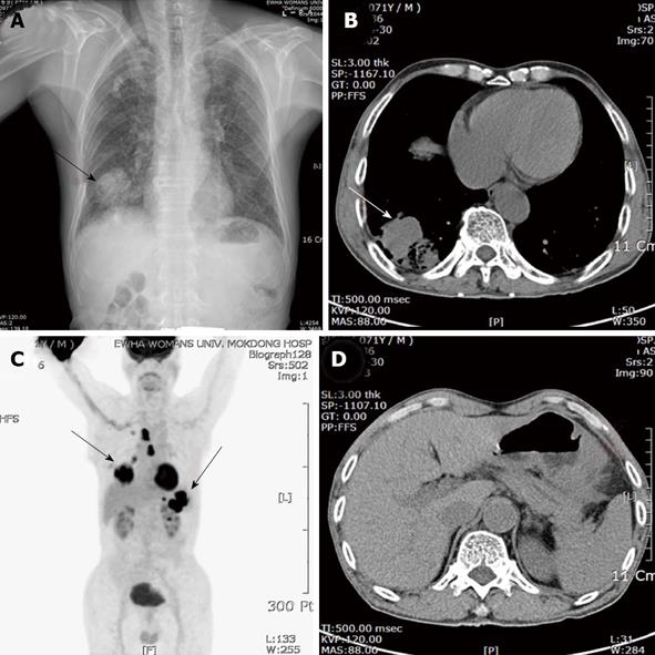
Figure 1 Chest posteroanterior view, computed tomography scan and (18F) fluoro-2-deoxy-D-glucose positron emission tomography-computed tomography.
A: A solitary mass with solid opacity in the right lower lung (RLL) (arrow); B: Chest computed tomography (CT) scan demonstrated an irregular mass measuring 5.8 cm in size with central necrosis at RLL (arrow); C: [18F] fluoro-2-deoxy-D-glucose-positron emission tomography (FDG-PET)/CT showed a FDG-avid mass on the right lower lobe, mediastinum, stomach and splenic hilum; D: Abdominal CT showed an encircling mass at the body of the stomach with multiple enlarged perigastric lymph nodes and splenic hilar invasion.
- Citation: Kim YI, Kang BC, Sung SH. Surgically resected gastric metastasis of pulmonary squamous cell carcinoma. World J Gastrointest Surg 2013; 5(10): 278-281
- URL: https://www.wjgnet.com/1948-9366/full/v5/i10/278.htm
- DOI: https://dx.doi.org/10.4240/wjgs.v5.i10.278


