Copyright
©2012 Baishideng.
World J Gastrointest Surg. Jun 27, 2012; 4(6): 157-162
Published online Jun 27, 2012. doi: 10.4240/wjgs.v4.i6.157
Published online Jun 27, 2012. doi: 10.4240/wjgs.v4.i6.157
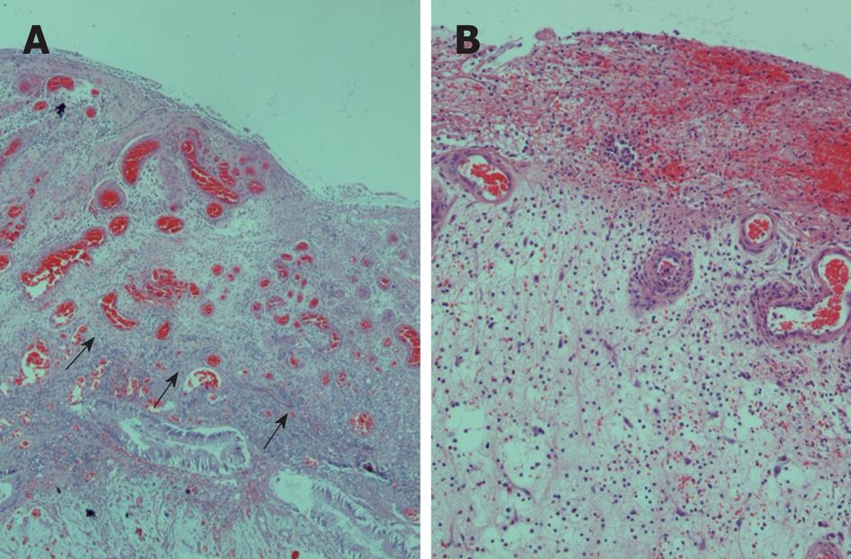
Figure 2 A cap of inflamed and ulcerated granulation tissue was observed in most superficial regions (arrows) (A), covered by fibrin and inflammatory exudate, as presented in higher magnification in (B).
- Citation: Papaconstantinou I, Karakatsanis A, Benia X, Polymeneas G, Kostopoulou E. Solitary rectal cap polyp: Case report and review of the literature. World J Gastrointest Surg 2012; 4(6): 157-162
- URL: https://www.wjgnet.com/1948-9366/full/v4/i6/157.htm
- DOI: https://dx.doi.org/10.4240/wjgs.v4.i6.157


