Copyright
©2012 Baishideng.
World J Gastrointest Surg. May 27, 2012; 4(5): 104-113
Published online May 27, 2012. doi: 10.4240/wjgs.v4.i5.104
Published online May 27, 2012. doi: 10.4240/wjgs.v4.i5.104
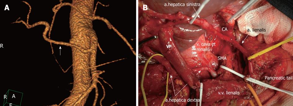
Figure 2 Michel’s type II celiac-mesenterial anatomy.
A: 3D computed tomography angiographic image. Replaced right hepatic artery (white arrow); B: View of the operating field after the extended pancreaticoduodenectomy. Note a the presence of a replaced right hepatic artery originating from the superior mesenteric artery (SMA). CA: Celiac artery.
- Citation: Zakharova OP, Karmazanovsky GG, Egorov VI. Pancreatic adenocarcinoma: Outstanding problems. World J Gastrointest Surg 2012; 4(5): 104-113
- URL: https://www.wjgnet.com/1948-9366/full/v4/i5/104.htm
- DOI: https://dx.doi.org/10.4240/wjgs.v4.i5.104


