Copyright
©2012 Baishideng Publishing Group Co.
World J Gastrointest Surg. Mar 27, 2012; 4(3): 79-82
Published online Mar 27, 2012. doi: 10.4240/wjgs.v4.i3.79
Published online Mar 27, 2012. doi: 10.4240/wjgs.v4.i3.79
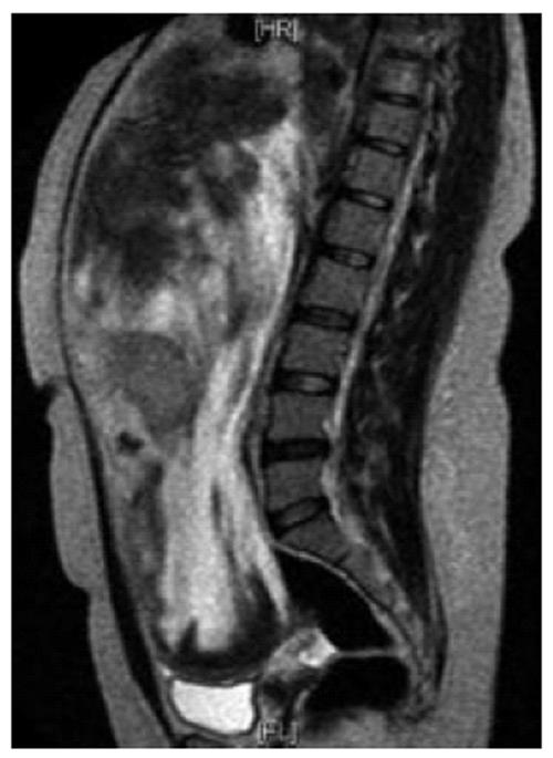
Figure 2 Magnetic resonance imaging abdomen and pelvis.
T2 weighted image (sagittal section) showing a huge intraperitoneal well defined mass displacing the liver and bowels cranially and bladder caudally with heterogenous bright and and low signals.
- Citation: Gari MKM, Guraya SY, Hussein AM, Hego MMN. Giant mesenteric fibromatosis: Report of a case and review of the literature. World J Gastrointest Surg 2012; 4(3): 79-82
- URL: https://www.wjgnet.com/1948-9366/full/v4/i3/79.htm
- DOI: https://dx.doi.org/10.4240/wjgs.v4.i3.79


