Copyright
©2012 Baishideng Publishing Group Co.
World J Gastrointest Surg. Mar 27, 2012; 4(3): 73-78
Published online Mar 27, 2012. doi: 10.4240/wjgs.v4.i3.73
Published online Mar 27, 2012. doi: 10.4240/wjgs.v4.i3.73
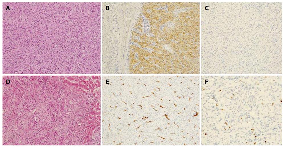
Figure 6 Microscopic appearance of the lesion.
A: Histological examination revealed a tumor without fibrous capsule with interlacing bands of uniform spindle cells whose elongated nuclei were arranged in a palisading pattern (hematoxylin-eosin, original magnification × 200); B: Border area between tumor and non-tumor; C-E: Immunohistochemical analysis revealed that tumor cells were diffusely and strongly positive for S-100 (C), but negative for c-kit (D), or CD34 (E); F: The Ki-67 labeling index was 2%.
- Citation: Hayashi M, Takeshita A, Yamamoto K, Tanigawa N. Primary hepatic benign schwannoma. World J Gastrointest Surg 2012; 4(3): 73-78
- URL: https://www.wjgnet.com/1948-9366/full/v4/i3/73.htm
- DOI: https://dx.doi.org/10.4240/wjgs.v4.i3.73


