Copyright
©2012 Baishideng Publishing Group Co.
World J Gastrointest Surg. Mar 27, 2012; 4(3): 73-78
Published online Mar 27, 2012. doi: 10.4240/wjgs.v4.i3.73
Published online Mar 27, 2012. doi: 10.4240/wjgs.v4.i3.73
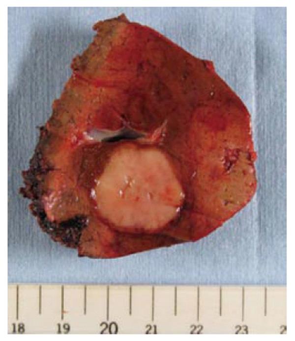
Figure 5 Macroscopic appearance of the lesion.
Macroscopically, the resected specimen showed a tumor of 23 mm in diameter. The cut section showed a solid component. The tumor had no cystic component and lacked apparent capsule.
- Citation: Hayashi M, Takeshita A, Yamamoto K, Tanigawa N. Primary hepatic benign schwannoma. World J Gastrointest Surg 2012; 4(3): 73-78
- URL: https://www.wjgnet.com/1948-9366/full/v4/i3/73.htm
- DOI: https://dx.doi.org/10.4240/wjgs.v4.i3.73


