Copyright
©2012 Baishideng Publishing Group Co.
World J Gastrointest Surg. Jan 27, 2012; 4(1): 1-8
Published online Jan 27, 2012. doi: 10.4240/wjgs.v4.i1.1
Published online Jan 27, 2012. doi: 10.4240/wjgs.v4.i1.1
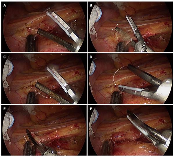
Figure 4 Skeletonization technique for the feeding artery.
A: First, the ultrasonic surgical blade of the Harmonic ACE penetrates the sheath of the feeding artery; B: The dissection margin is enlarged by moving the surgical blade along the feeding artery; C: After sufficient enlargement of the dissection margin for the sheath of the feeding artery, the ultrasonic surgical blade of the Harmonic ACE is removed from the sheath of the feeding artery; D: Next, the tip of the Harmonic ACE is rotated; E: The tissue pad side of the Harmonic ACE is applied in the penetrated space of the sheath of the feeding artery; F: One side of the sheath of the feeding artery is sealed and skeletonized safely. Then, the other side of the sheath is treated similarly, resulting in the complete skeletonization of the feeding artery.
- Citation: Hotta T, Takifuji K, Yokoyama S, Matsuda K, Higashiguchi T, Tominaga T, Oku Y, Watanabe T, Nasu T, Hashimoto T, Tamura K, Ieda J, Yamamoto N, Iwamoto H, Yamaue H. Literature review of the energy sources for performing laparoscopic colorectal surgery. World J Gastrointest Surg 2012; 4(1): 1-8
- URL: https://www.wjgnet.com/1948-9366/full/v4/i1/1.htm
- DOI: https://dx.doi.org/10.4240/wjgs.v4.i1.1


