Copyright
©2011 Baishideng Publishing Group Co.
World J Gastrointest Surg. Apr 27, 2011; 3(4): 56-58
Published online Apr 27, 2011. doi: 10.4240/wjgs.v3.i4.56
Published online Apr 27, 2011. doi: 10.4240/wjgs.v3.i4.56
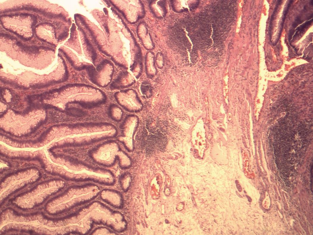
Figure 4 Histology picture, showing the tubular and villous component of the adenoma and the non-invaded muscularis mucosa (HE stain).
- Citation: Dardamanis D, Theodorou D, Theodoropoulos G, Larentzakis A, Natoudi M, Doulami G, Zoumpouli C, Markogiannakis H, Katsaragakis S, Zografos GC. Transanal polypectomy using single incision laparoscopic instruments. World J Gastrointest Surg 2011; 3(4): 56-58
- URL: https://www.wjgnet.com/1948-9366/full/v3/i4/56.htm
- DOI: https://dx.doi.org/10.4240/wjgs.v3.i4.56


