Copyright
©2011 Baishideng Publishing Group Co.
World J Gastrointest Surg. Mar 27, 2011; 3(3): 31-38
Published online Mar 27, 2011. doi: 10.4240/wjgs.v3.i3.31
Published online Mar 27, 2011. doi: 10.4240/wjgs.v3.i3.31
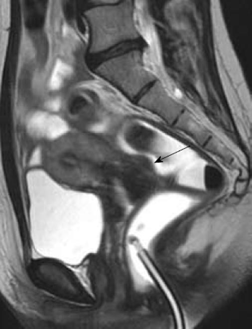
Figure 4 Magnetic resonance imaging enteroclysis.
The rectosigmoid is distended by using 250-300 mL of ultrasonographic gel diluted with saline solution; the 20-Fr Foley catheter used for retrograde distension can be observed in the figure. The fluid solution has a biphasic behavior on MR sequences: hypointense in T1W images and hyperintense in T2W images. A small rectovaginal endometriotic nodule (larger diameter 12 mm) is observed (arrow).
- Citation: Ferrero S, Camerini G, Maggiore ULR, Venturini PL, Biscaldi E, Remorgida V. Bowel endometriosis: Recent insights and unsolved problems. World J Gastrointest Surg 2011; 3(3): 31-38
- URL: https://www.wjgnet.com/1948-9366/full/v3/i3/31.htm
- DOI: https://dx.doi.org/10.4240/wjgs.v3.i3.31


