Copyright
©2011 Baishideng Publishing Group Co.
World J Gastrointest Surg. Jan 27, 2011; 3(1): 16-20
Published online Jan 27, 2011. doi: 10.4240/wjgs.v3.i1.16
Published online Jan 27, 2011. doi: 10.4240/wjgs.v3.i1.16
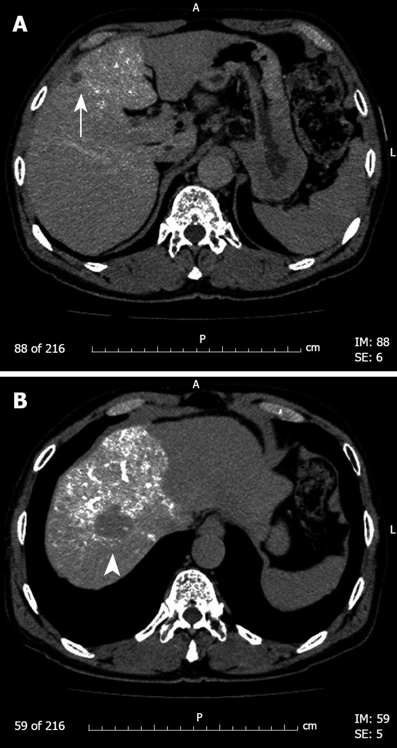
Figure 1 Preoperative abdominal computed tomography during angiography.
This computed tomography reveals low density areas indicating two hepatocellular carcinoma nodules (arrow, arrowhead) of about 1.3 and 3 cm in diameter, in liver segments 8 (A) and 4 (B).
- Citation: Inoue Y, Hayashi M, Hirokawa F, Takeshita A, Tanigawa N. Peritoneovenous shunt for intractable ascites due to hepatic lymphorrhea after hepatectomy. World J Gastrointest Surg 2011; 3(1): 16-20
- URL: https://www.wjgnet.com/1948-9366/full/v3/i1/16.htm
- DOI: https://dx.doi.org/10.4240/wjgs.v3.i1.16


