Copyright
©2010 Baishideng Publishing Group Co.
World J Gastrointest Surg. Aug 27, 2010; 2(8): 260-264
Published online Aug 27, 2010. doi: 10.4240/wjgs.v2.i8.260
Published online Aug 27, 2010. doi: 10.4240/wjgs.v2.i8.260
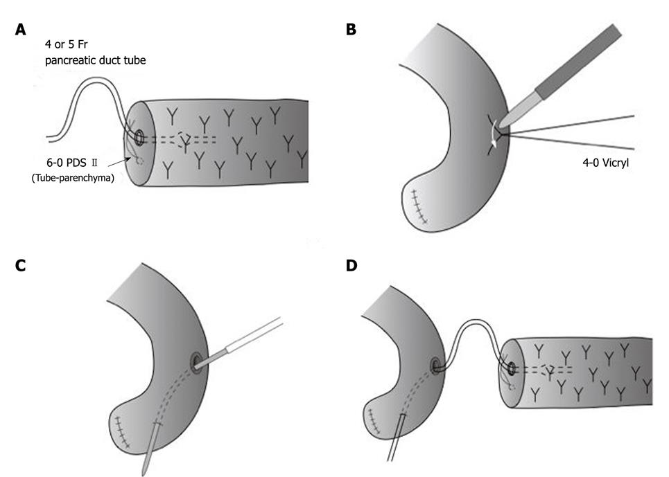
Figure 1 Preparation of pancreaticojejunostomy using pancreatic duct tube.
A: A pancreatic duct tube is inserted as a stent tube into the main pancreatic duct of the pancreatic remnant from the cut-end; B: A very small part (usually 2-3 mm in diameter) of the jejunal serosa is cut by an electric cautery; C: The metallic needle of the pancreatic duct tube is introduced into the jejunum through the site of serosal cut and then taken out of the distal end of the jejunum (Roux-en-Y jejunal limb); D: The distance between the induced jejunal hole and the cut end of the main pancreatic duct should be kept 5 cm away or more, putting the pancreatic duct tube like a long loop during pancreaticojejunostomy.
- Citation: Azumi Y, Isaji S, Kato H, Nobuoka Y, Kuriyama N, Kishiwada M, Hamada T, Mizuno S, Usui M, Sakurai H, Tabata M. A standardized technique for safe pancreaticojejunostomy: Pair-Watch suturing technique. World J Gastrointest Surg 2010; 2(8): 260-264
- URL: https://www.wjgnet.com/1948-9366/full/v2/i8/260.htm
- DOI: https://dx.doi.org/10.4240/wjgs.v2.i8.260


