Copyright
©2010 Baishideng.
World J Gastrointest Surg. Jul 27, 2010; 2(7): 247-250
Published online Jul 27, 2010. doi: 10.4240/wjgs.v2.i7.247
Published online Jul 27, 2010. doi: 10.4240/wjgs.v2.i7.247
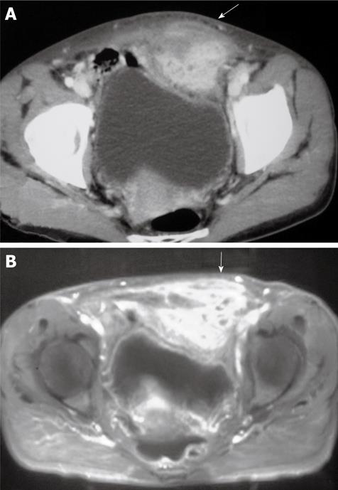
Figure 1 Diagnostic images of the patient.
A: Computed tomography scan shows a 6 cm × 5 cm solid mass that originated from the left inferior portion of the rectus muscle of the abdominal wall (arrow); B: Magnetic resonance image confirms that the solid mass originated from the left inferior portion of the rectus muscle of the abdominal wall (arrow).
- Citation: Acquaro P, Tagliabue F, Confalonieri G, Faccioli P, Costa M. Abdominal wall actinomycosis simulating a malignant neoplasm: Case report and review of the literature. World J Gastrointest Surg 2010; 2(7): 247-250
- URL: https://www.wjgnet.com/1948-9366/full/v2/i7/247.htm
- DOI: https://dx.doi.org/10.4240/wjgs.v2.i7.247


