Copyright
©2010 Baishideng Publishing Group Co.
World J Gastrointest Surg. Dec 27, 2010; 2(12): 405-408
Published online Dec 27, 2010. doi: 10.4240/wjgs.v2.i12.405
Published online Dec 27, 2010. doi: 10.4240/wjgs.v2.i12.405
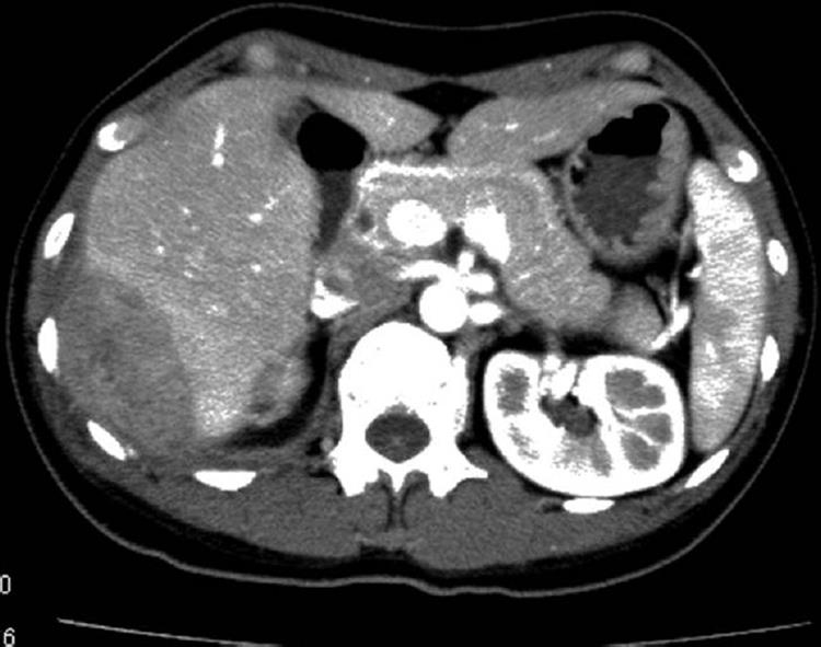
Figure 3 Preoperative computed tomography image of case 2.
Tumor 70 mm × 45 mm in the right flank is observed invading to segment 6 of the liver, showing an irregularly circumferentiated, contrast-enhanced cystic structure with heterogenous content.
- Citation: Hayashi M, Asakuma M, Tsunemi S, Inoue Y, Shimizu T, Komeda K, Hirokawa F, Takeshita A, Egashira Y, Tanigawa N. Surgical treatment for abdominal actinomycosis: A report of two cases. World J Gastrointest Surg 2010; 2(12): 405-408
- URL: https://www.wjgnet.com/1948-9366/full/v2/i12/405.htm
- DOI: https://dx.doi.org/10.4240/wjgs.v2.i12.405


