Copyright
©2010 Baishideng Publishing Group Co.
World J Gastrointest Surg. Dec 27, 2010; 2(12): 405-408
Published online Dec 27, 2010. doi: 10.4240/wjgs.v2.i12.405
Published online Dec 27, 2010. doi: 10.4240/wjgs.v2.i12.405
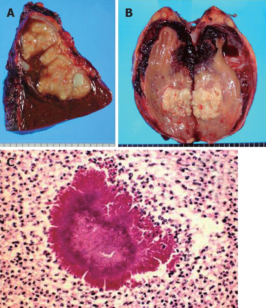
Figure 2 Resected liver (A), resected kidney (B) and pathological examination of the resected kidney (C) of case 1.
A: The lesion in the resected liver was a macroscopically whitish node with capsule; B: The resected kidney showed the same nature as the liver tumor; C: The pathological examination of the resected kidney revealed a typical abscess due to Actinomyces israelii. Note the abscess contained filamentous bacterial colonies, i.e. sulfur granules.
- Citation: Hayashi M, Asakuma M, Tsunemi S, Inoue Y, Shimizu T, Komeda K, Hirokawa F, Takeshita A, Egashira Y, Tanigawa N. Surgical treatment for abdominal actinomycosis: A report of two cases. World J Gastrointest Surg 2010; 2(12): 405-408
- URL: https://www.wjgnet.com/1948-9366/full/v2/i12/405.htm
- DOI: https://dx.doi.org/10.4240/wjgs.v2.i12.405


