Copyright
©2010 Baishideng Publishing Group Co.
World J Gastrointest Surg. Oct 27, 2010; 2(10): 337-341
Published online Oct 27, 2010. doi: 10.4240/wjgs.v2.i10.337
Published online Oct 27, 2010. doi: 10.4240/wjgs.v2.i10.337
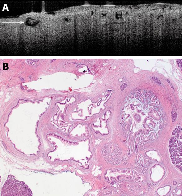
Figure 2 An optical coherence tomography image of a patient with borderline intraductal papillary mucinous neoplasm (A) and photomicrograph of an intraductal papillary mucinous adenoma in the same patient (B).
A: Multiple cystic lesions are pictured here with medium to high scattering in the cyst cavity; the scattering suggests the existence of mucin. A single, mucinous cystic lesion is indicated by the white arrow and scattering is clearly seen within the cystic structure. Image provided courtesy of Dr. Sevde Cizginer at Massachusetts General Hospital, Boston, MA: B: Photomicrograph of an intraductal papillary mucinous adenoma in the same patient. The cysts are lined by a single layer of foveolar-type epithelial cells. Focally, papillary areas are identified. Image provided courtesy of Dr. Vikram Deshpande at Massachusetts General Hospital, Boston, MA.
- Citation: Turner BG, Brugge WR. Diagnostic and therapeutic endoscopic approaches to intraductal papillary mucinous neoplasm. World J Gastrointest Surg 2010; 2(10): 337-341
- URL: https://www.wjgnet.com/1948-9366/full/v2/i10/337.htm
- DOI: https://dx.doi.org/10.4240/wjgs.v2.i10.337


