Copyright
©2010 Baishideng Publishing Group Co.
World J Gastrointest Surg. Oct 27, 2010; 2(10): 324-330
Published online Oct 27, 2010. doi: 10.4240/wjgs.v2.i10.324
Published online Oct 27, 2010. doi: 10.4240/wjgs.v2.i10.324
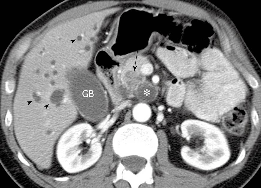
Figure 3 Axial contrast enhanced computed tomography image at the level of the head of the pancreas shows a cystic lesion (asterisk) in the uncinate process of the pancreas with a hypoattenuating area (arrow) in the adjacent pancreatic parenchyma.
Note the intrahepatic biliary dilatation (arrowheads) due to obstruction of the common bile duct (not shown) by the infiltrating mass. Invasive pancreatic adenocarcinoma arising from an intraductal papillary mucinous neoplasm was confirmed at pathology after a Whipple procedure. GB: Gallbladder.
- Citation: Pedrosa I, Boparai D. Imaging considerations in intraductal papillary mucinous neoplasms of the pancreas. World J Gastrointest Surg 2010; 2(10): 324-330
- URL: https://www.wjgnet.com/1948-9366/full/v2/i10/324.htm
- DOI: https://dx.doi.org/10.4240/wjgs.v2.i10.324


