Copyright
©2010 Baishideng Publishing Group Co.
World J Gastrointest Surg. Oct 27, 2010; 2(10): 324-330
Published online Oct 27, 2010. doi: 10.4240/wjgs.v2.i10.324
Published online Oct 27, 2010. doi: 10.4240/wjgs.v2.i10.324
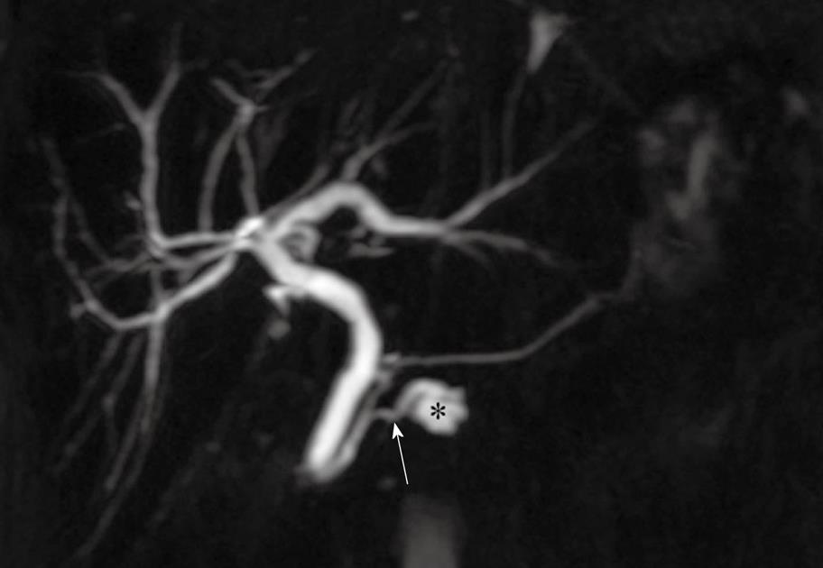
Figure 1 Coronal maximum intensity projection from a 3D T2-weighted MRCP acquisition shows a cystic lesion in the uncinate process of the pancreas (asterisk) and a communicating branch duct (arrow) between the cyst and the normal caliber main pancreatic duct.
These findings are characteristic of a branch duct intraductal papillary mucinous neoplasm and this lesion has been stable on follow up MRCP examinations for 3 years.
- Citation: Pedrosa I, Boparai D. Imaging considerations in intraductal papillary mucinous neoplasms of the pancreas. World J Gastrointest Surg 2010; 2(10): 324-330
- URL: https://www.wjgnet.com/1948-9366/full/v2/i10/324.htm
- DOI: https://dx.doi.org/10.4240/wjgs.v2.i10.324


