Copyright
©2010 Baishideng Publishing Group Co.
World J Gastrointest Surg. Oct 27, 2010; 2(10): 306-313
Published online Oct 27, 2010. doi: 10.4240/wjgs.v2.i10.306
Published online Oct 27, 2010. doi: 10.4240/wjgs.v2.i10.306
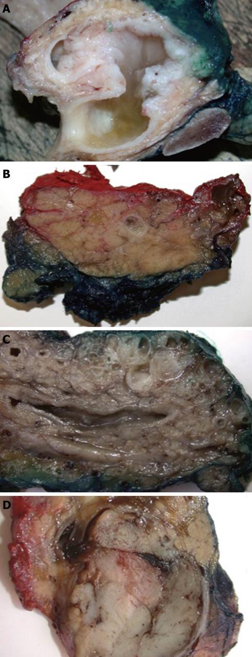
Figure 1 Variation in gross appearance of intraductal papillary-mucinous neoplasm.
A: Intraductal papillary-mucinous neoplasm (IPMN) involving the main pancreatic duct with copious mucus and prominent intraductal tumor proliferation causing marked dilatation of the Wirsung and Santorini ducts; B: IPMN of the main duct characterized by subtle granularity of the duct wall, little grossly visible mucin and minimal duct dilatation; C: IPMN involving clusters of dilated and mucin-filled branched ducts. Note mild distension and mucinous content of the main pancreatic duct; D: Distension of a branch duct by solid tumor tissue of an oncocytic-type IPMN.
- Citation: Verbeke CS. Intraductal papillary-mucinous neoplasia of the pancreas: Histopathology and molecular biology. World J Gastrointest Surg 2010; 2(10): 306-313
- URL: https://www.wjgnet.com/1948-9366/full/v2/i10/306.htm
- DOI: https://dx.doi.org/10.4240/wjgs.v2.i10.306


