Copyright
©The Author(s) 2025.
World J Gastrointest Surg. Apr 27, 2025; 17(4): 103494
Published online Apr 27, 2025. doi: 10.4240/wjgs.v17.i4.103494
Published online Apr 27, 2025. doi: 10.4240/wjgs.v17.i4.103494
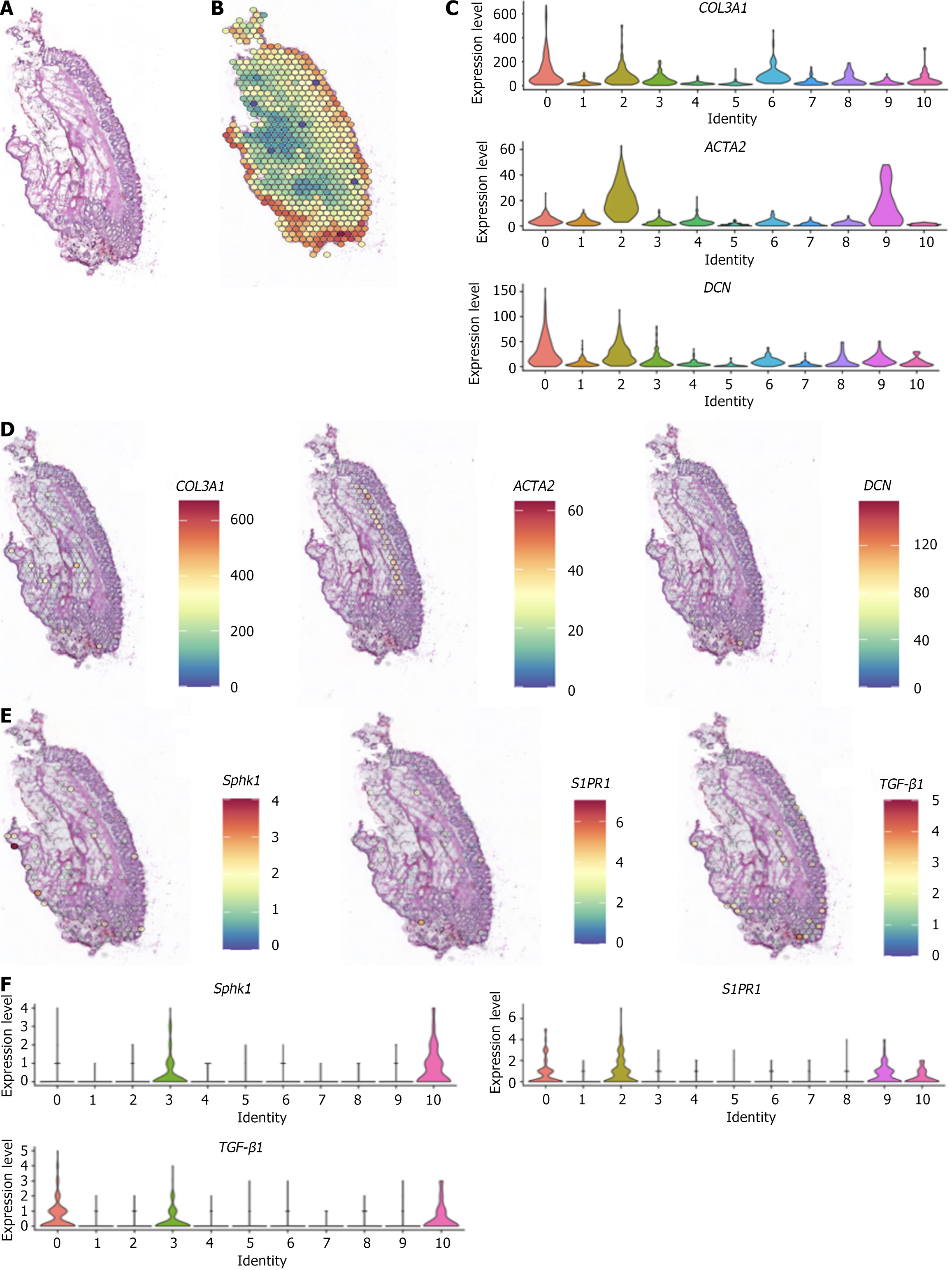
Figure 2 Spatial transcriptome map of internal hemorrhoid tissue.
A: Hematoxylin and eosin-stained section of internal hemorrhoid tissue; B: Spatial transcriptome; C: Differences in expression of the classic marker genes collagen 3 alpha 1, decorin, and actin alpha 2 in fibroblasts; D: Spatial localization of the classic marker genes collagen 3 alpha 1, decorin, and actin alpha 2 in fibroblasts; E: Spatial distribution of sphingosine kinase 1, sphingosine-1-phosphate receptor 1, and transforming growth factor-β1; F: Differences in gene expression of sphingosine kinase 1, sphingosine-1-phosphate receptor 1, and transforming growth factor-β1. COL3A1: Collagen 3 alpha 1; DCN: Decorin; ACTA2: Actin alpha 2; Sphk1: Sphingosine kinase 1; S1PR1: Sphingosine-1-phosphate receptor 1; TGF-β1: Transforming growth factor-β1.
- Citation: Ke MH, Huang SY, Lin WG, Xu ZG, Zheng XX, Liu XB, Cheng YM, Li ZF. Single-nucleus RNA sequencing and spatial transcriptomics reveal the mechanism by which Xiaozhiling injection treats internal hemorrhoids. World J Gastrointest Surg 2025; 17(4): 103494
- URL: https://www.wjgnet.com/1948-9366/full/v17/i4/103494.htm
- DOI: https://dx.doi.org/10.4240/wjgs.v17.i4.103494


