Copyright
©The Author(s) 2025.
World J Gastrointest Surg. Apr 27, 2025; 17(4): 102730
Published online Apr 27, 2025. doi: 10.4240/wjgs.v17.i4.102730
Published online Apr 27, 2025. doi: 10.4240/wjgs.v17.i4.102730
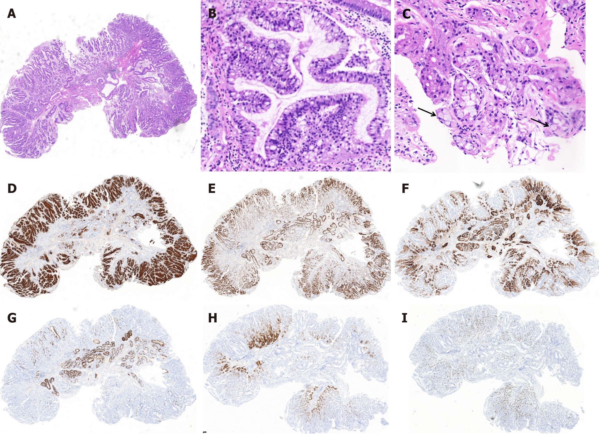
Figure 2 Representative hematoxylin and eosin staining and immunohistochemical analysis images from a case involving a middle-aged male patient diagnosed with gastric adenocarcinoma of the fundic gland mucosa.
A: The superficial mucosa exhibited gastric foveolar-type epithelium, numerous mucous glands were observed immediately below the epithelium; B: Scattered dilated glands were observed in the deepest region of the mucosa; C: Focal areas displayed several signet-ring cell changes (black arrow); D-I: Gastric foveolar-type epithelium was diffusely positivity for MUC5AC (D) and Ki67 (E); mucous glands were positivity for MUC6 (F), partially positive for MUC2 (G), pepsinogen I (H) and H+/K+ ATPase (I).
- Citation: Yang QY, Xu J, Hu JW, Huang XD. Gastric adenocarcinoma of fundic gland mucosa arising in heterotopic gastric mucosa of the duodenum: A case report. World J Gastrointest Surg 2025; 17(4): 102730
- URL: https://www.wjgnet.com/1948-9366/full/v17/i4/102730.htm
- DOI: https://dx.doi.org/10.4240/wjgs.v17.i4.102730


