Copyright
©The Author(s) 2025.
World J Gastrointest Surg. Mar 27, 2025; 17(3): 102428
Published online Mar 27, 2025. doi: 10.4240/wjgs.v17.i3.102428
Published online Mar 27, 2025. doi: 10.4240/wjgs.v17.i3.102428
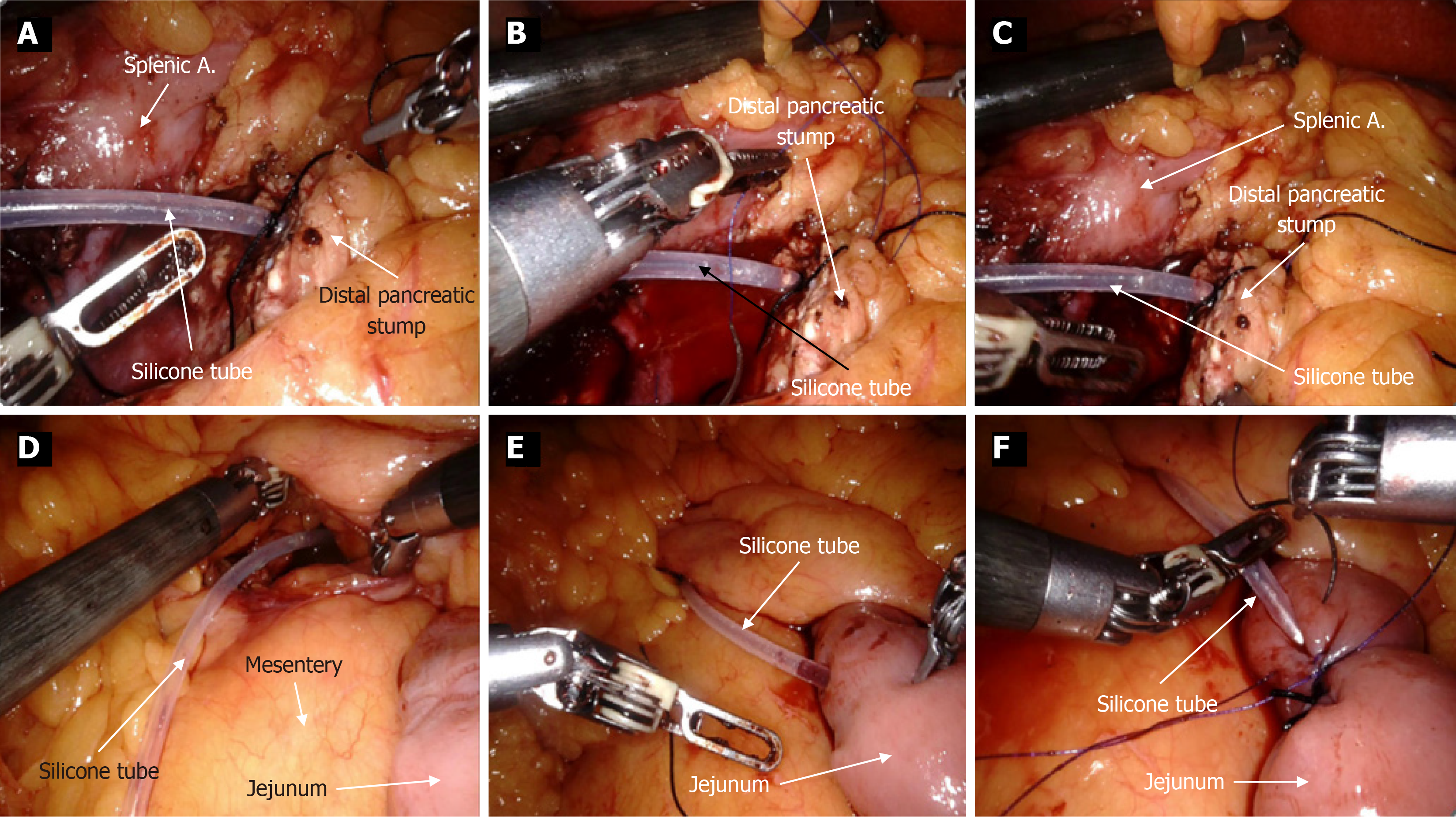
Figure 3 Clinical photographs depicting the step-by-step process of the bridge drainage procedure.
A: Tie a suture around the surface of the drainage tube and then insert it into the main pancreatic duct; B: Use 4-0 polypropylene non-absorbable suture to create a U-shaped stitch; C: Secure the Prolene suture and the tied suture together in an interlocking manner; D: Pass the other side of the drainage tube through the transverse mesocolon; E: Pass through the transverse mesocolon and into the jejunum approximately 10-15 cm below the ligament of Treitz; F: Secure and embed the drainage tube using barbed sutures. A: Artery.
- Citation: Lu XY, Tan XD. Clinical outcomes of interlocking main pancreatic duct-jejunal internal bridge drainage in middle pancreatectomy: A comparative study. World J Gastrointest Surg 2025; 17(3): 102428
- URL: https://www.wjgnet.com/1948-9366/full/v17/i3/102428.htm
- DOI: https://dx.doi.org/10.4240/wjgs.v17.i3.102428


