Copyright
©The Author(s) 2025.
World J Gastrointest Surg. Mar 27, 2025; 17(3): 102174
Published online Mar 27, 2025. doi: 10.4240/wjgs.v17.i3.102174
Published online Mar 27, 2025. doi: 10.4240/wjgs.v17.i3.102174
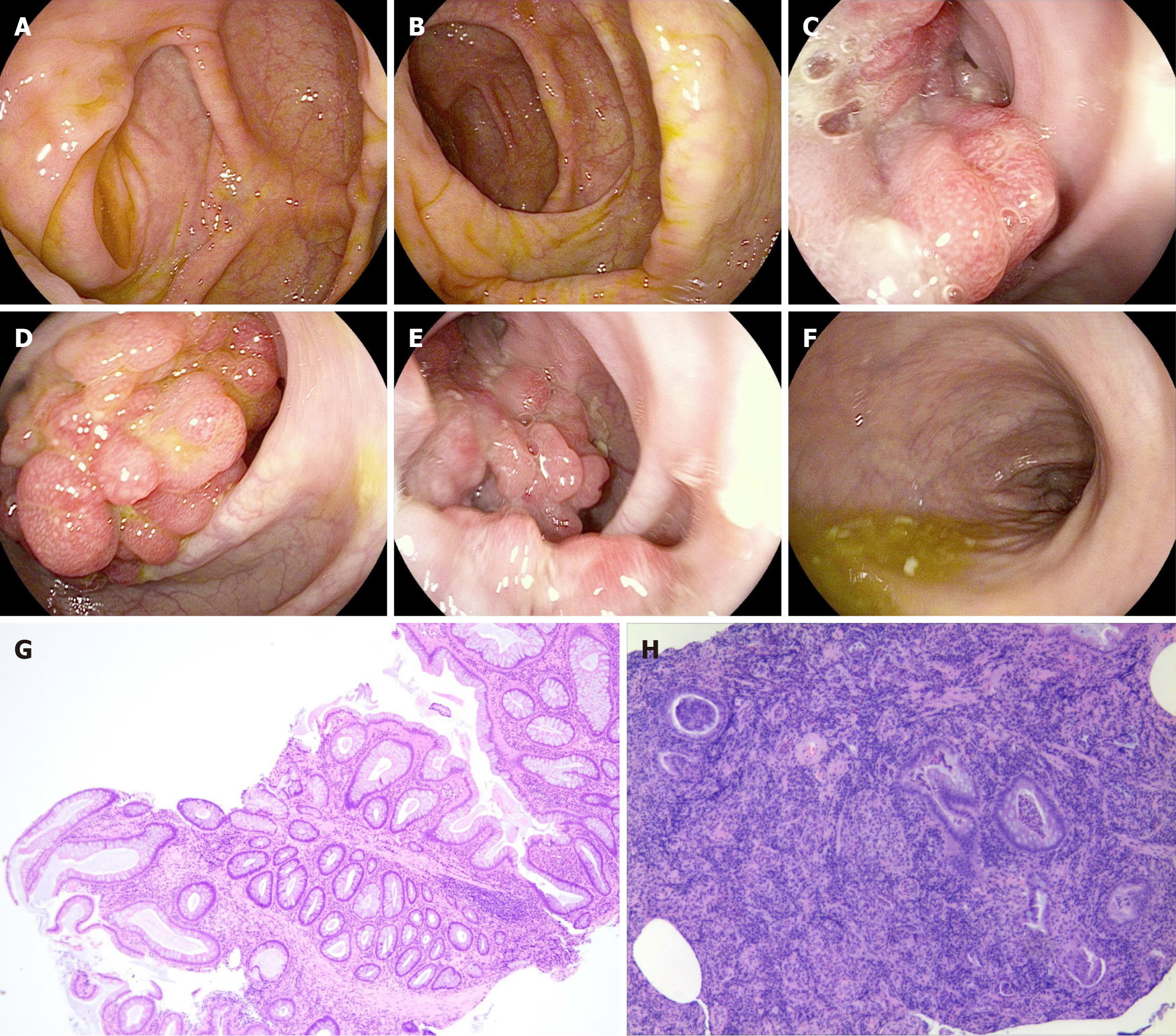
Figure 4 Colonoscopy and endoscopic forceps were used to obtain tissue pathological biopsy.
A: The appendiceal fossa mucosa was smooth without dysplasia or polyps; B: The ileocecal mucosa was smooth and no ulcer or neoplasm was observed; C-E: A grape-like mass was seen in the sigmoid colon 30 cm to 35 cm from the anal verge; F: No abnormal lesions were found in the rectum; G: Pathological changes of hamartomatous polyps; H: Pathological examination showed mucosal prolapsed changes and ulcer formation in some areas.
- Citation: Tian ZS, Ma XP, Ruan HX, Yang Y, Zhao YL. Rare large sigmoid hamartomatous polyp in an elderly patient with atypical Peutz-Jeghers syndrome: A case report. World J Gastrointest Surg 2025; 17(3): 102174
- URL: https://www.wjgnet.com/1948-9366/full/v17/i3/102174.htm
- DOI: https://dx.doi.org/10.4240/wjgs.v17.i3.102174


