Copyright
©The Author(s) 2025.
World J Gastrointest Surg. Mar 27, 2025; 17(3): 102174
Published online Mar 27, 2025. doi: 10.4240/wjgs.v17.i3.102174
Published online Mar 27, 2025. doi: 10.4240/wjgs.v17.i3.102174
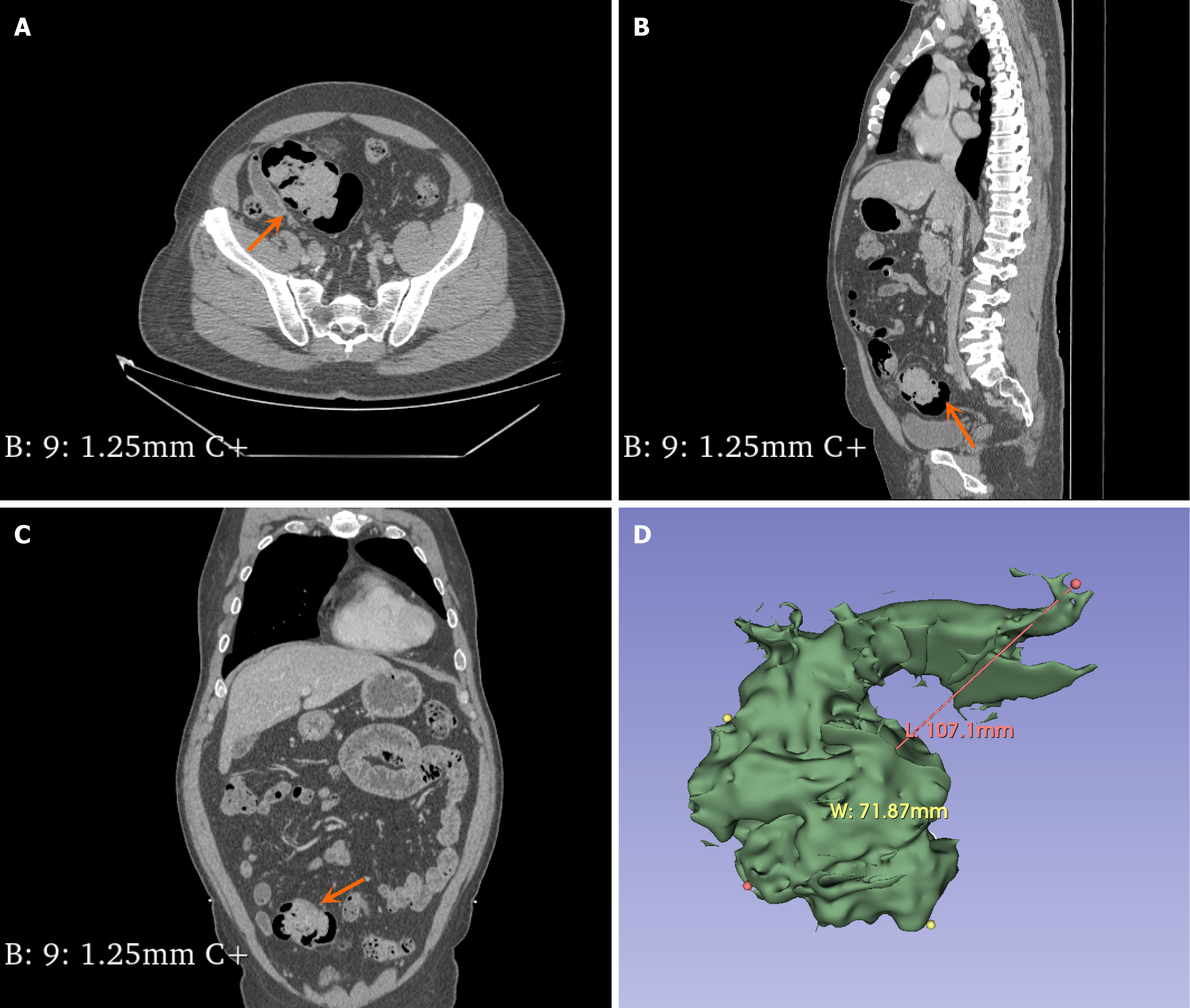
Figure 3 Contrast-enhanced computed tomography of the abdomen.
A: The sigmoid colon mass was observed in the horizontal plane; B: The sigmoid colon mass was observed in the sagittal planes; C: The sigmoid colon mass was observed in the coronal planes; D: 3D reconstruction model of sigmoid colon mass. The orange arrow points to the sigmoid mass.
- Citation: Tian ZS, Ma XP, Ruan HX, Yang Y, Zhao YL. Rare large sigmoid hamartomatous polyp in an elderly patient with atypical Peutz-Jeghers syndrome: A case report. World J Gastrointest Surg 2025; 17(3): 102174
- URL: https://www.wjgnet.com/1948-9366/full/v17/i3/102174.htm
- DOI: https://dx.doi.org/10.4240/wjgs.v17.i3.102174


