Copyright
©The Author(s) 2025.
World J Gastrointest Surg. Feb 27, 2025; 17(2): 97862
Published online Feb 27, 2025. doi: 10.4240/wjgs.v17.i2.97862
Published online Feb 27, 2025. doi: 10.4240/wjgs.v17.i2.97862
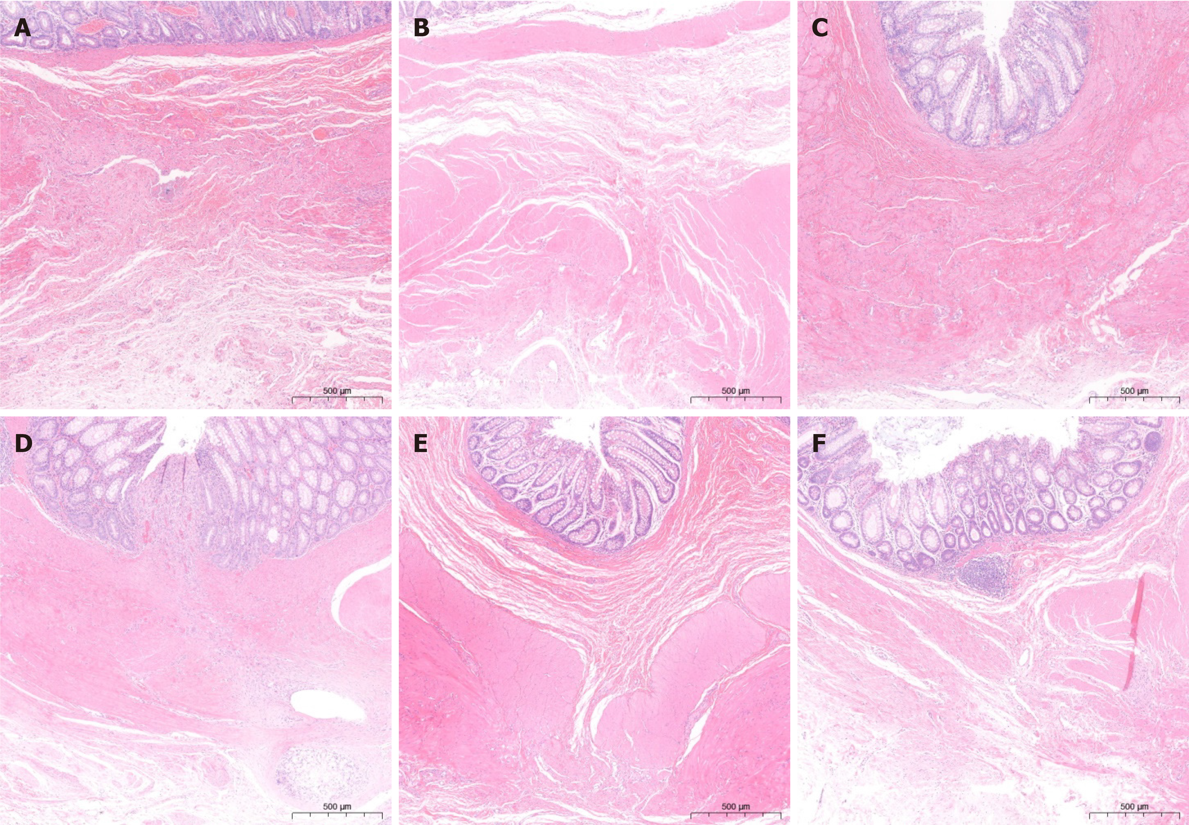
Figure 4 HE staining of the anastomosis specimens.
A: At one month after surgery, the anastomosis of the magnamosis group demonstrated an intact mucosa with minor inflammatory reactions and inadequate submucosal continuity; B: At 1-month postoperatively, the anastomosis of the suturing group showed a lack of mucosal and submucosal continuity, along with an increased inflammatory response; C: The anastomosis of the magnamosis group at 3 months postoperatively obviously improved; D: The anastomosis of the suturing anastomosis group at 3 months postoperatively also healed well, but the inflammatory responses remained; E: The anastomosis of the magnamosis group at 6 months postoperatively showed no inflammatory responses and well healing; F: The anastomosis of the suturing anastomosis group at 6 months postoperatively.
- Citation: Liu SQ, Zhang HK, Lv Y, Xu XH, Li YF, Quan DW. Magnamosis for rectal reconstruction in canines. World J Gastrointest Surg 2025; 17(2): 97862
- URL: https://www.wjgnet.com/1948-9366/full/v17/i2/97862.htm
- DOI: https://dx.doi.org/10.4240/wjgs.v17.i2.97862


