Copyright
©The Author(s) 2025.
World J Gastrointest Surg. Feb 27, 2025; 17(2): 97862
Published online Feb 27, 2025. doi: 10.4240/wjgs.v17.i2.97862
Published online Feb 27, 2025. doi: 10.4240/wjgs.v17.i2.97862
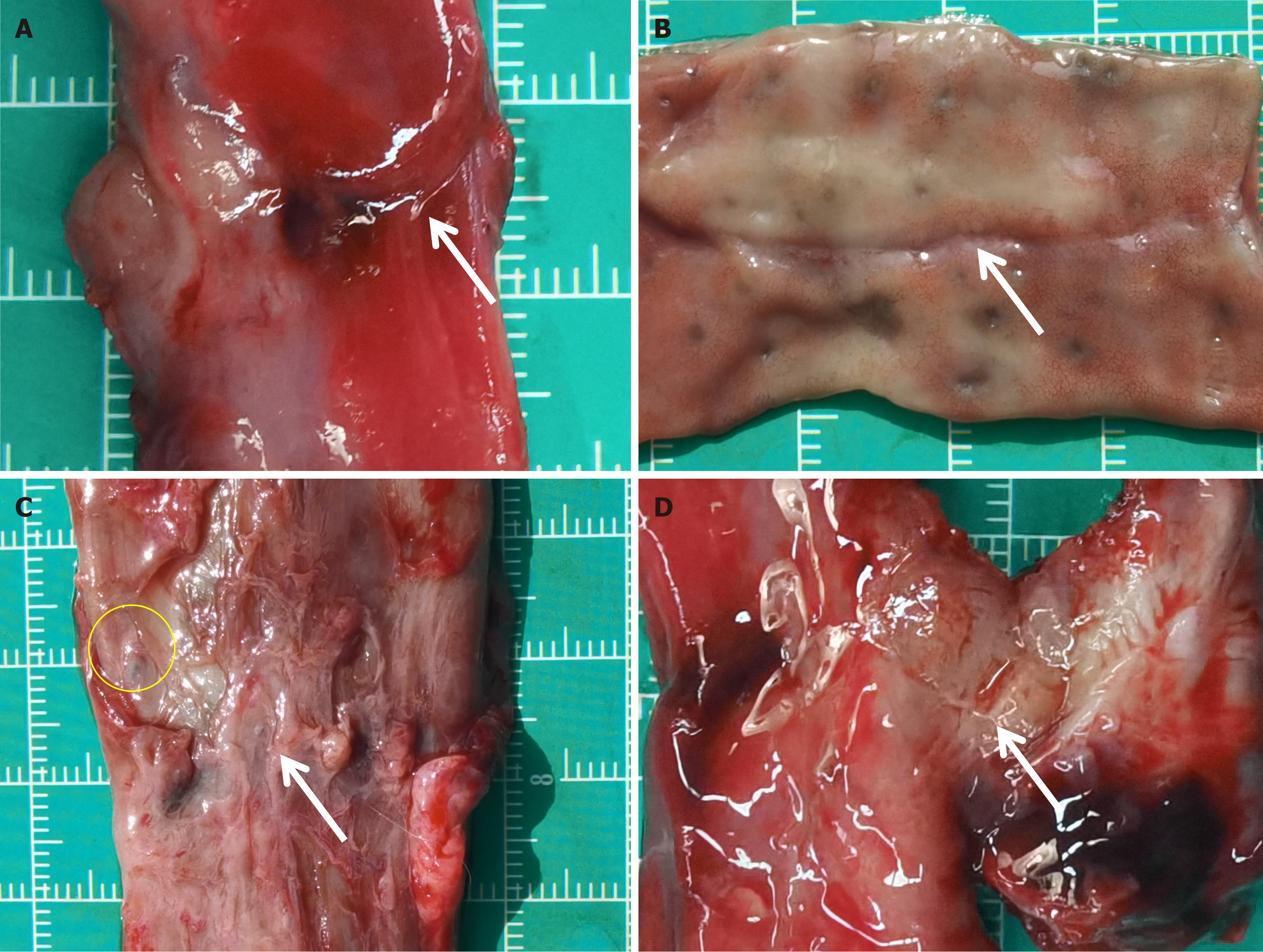
Figure 3 Gross observation of the anastomosis specimens.
A and B: The rectal anastomosis in the magnamosis group; C and D: The rectal anastomosis in the suturing anastomosis group. The white arrows indicate the anastomosis. The yellow circle shows the absorbable suture.
- Citation: Liu SQ, Zhang HK, Lv Y, Xu XH, Li YF, Quan DW. Magnamosis for rectal reconstruction in canines. World J Gastrointest Surg 2025; 17(2): 97862
- URL: https://www.wjgnet.com/1948-9366/full/v17/i2/97862.htm
- DOI: https://dx.doi.org/10.4240/wjgs.v17.i2.97862


