Copyright
©The Author(s) 2025.
World J Gastrointest Surg. Feb 27, 2025; 17(2): 94270
Published online Feb 27, 2025. doi: 10.4240/wjgs.v17.i2.94270
Published online Feb 27, 2025. doi: 10.4240/wjgs.v17.i2.94270
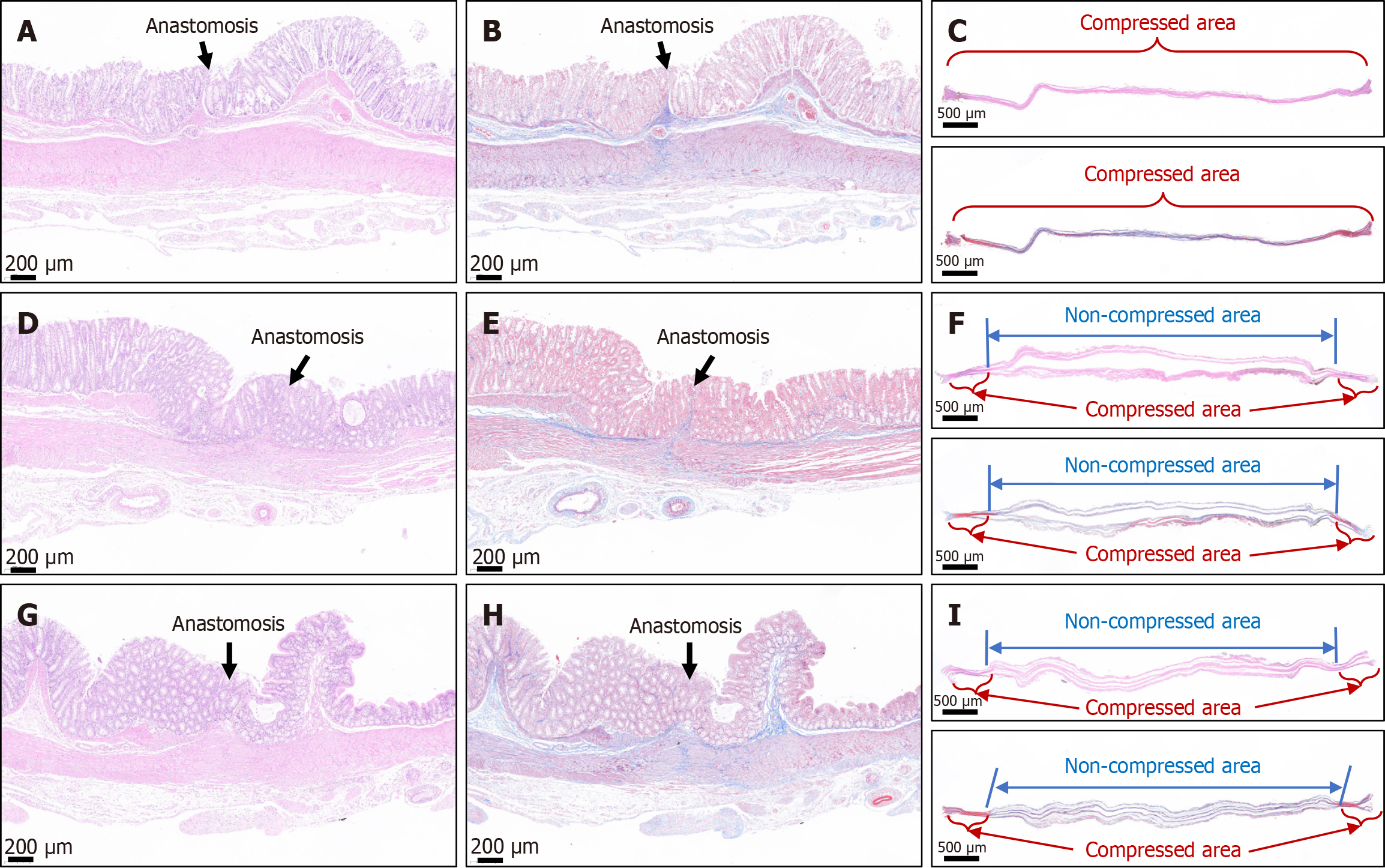
Figure 6 Representative images of histological staining of the colonic anastomosis.
A and B: Hematoxylin & eosin (H&E) and Masson trichrome staining of the anastomosis in the cylindrical group; C: H&E and Masson trichrome staining of the necrotic tissue between the daughter magnet (DM) and parent magnet (PM) in the cylindrical group; D and E: H&E and Masson trichrome staining of the anastomosis in the circular ring group; F: H&E and Masson trichrome staining of the necrotic tissue between the DM and PM in the circular ring group; G and H: H&E and Masson trichrome staining of the anastomosis in the cylindrical–circular ring group; I: H&E and Masson trichrome staining of the necrotic tissue between the DM and PM in the cylindrical–circular ring group.
- Citation: Zhang MM, Shi AH, Muensterer OJ, Uygun I, Lyu Y, Yan XP. Comparative study of cylindrical vs circular ring magnets for colonic anastomosis in rats. World J Gastrointest Surg 2025; 17(2): 94270
- URL: https://www.wjgnet.com/1948-9366/full/v17/i2/94270.htm
- DOI: https://dx.doi.org/10.4240/wjgs.v17.i2.94270


