Copyright
©The Author(s) 2024.
World J Gastrointest Surg. Sep 27, 2024; 16(9): 3065-3073
Published online Sep 27, 2024. doi: 10.4240/wjgs.v16.i9.3065
Published online Sep 27, 2024. doi: 10.4240/wjgs.v16.i9.3065
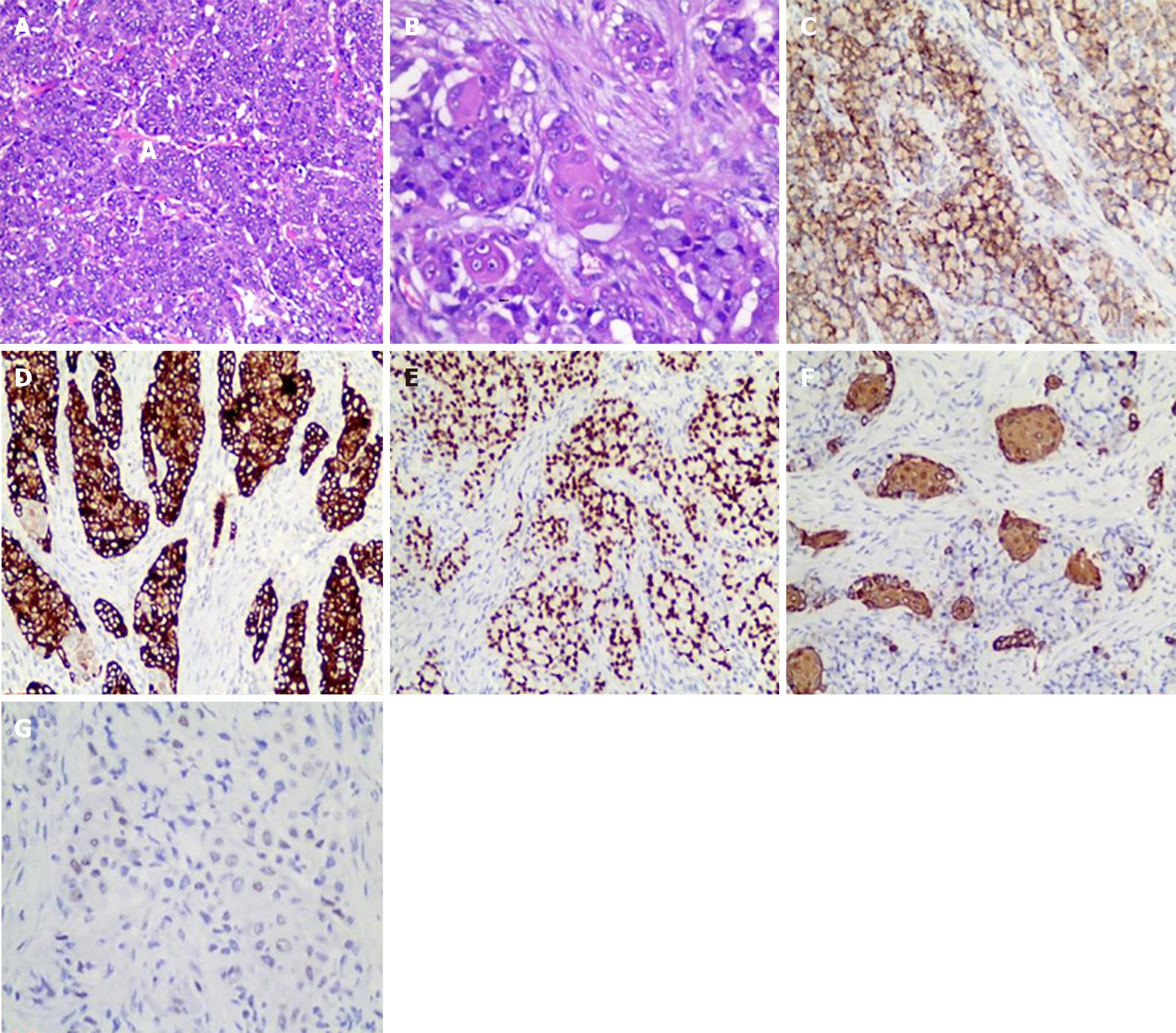
Figure 4 Histological examination and immunohistochemical staining results of the resected gastric specimens.
A: Adenocarcinoma components (magnification 100 ×); B: Squamous cell carcinoma components (magnification 200 ×); C: Novel aspartic proteinase A (magnification 100 ×); D: Cytokeratin 7 (magnification 100 ×); E: Thyroid transcription factor-1 (magnification 100 ×); F: Cytokeratin 5/6 (magnification 200 ×); G: P40 (magnification 200 ×).
- Citation: Lin Y, Wu YL, Zou DD, Luo XL, Zhang SY. Combined gastroscopic and laparoscopic resection of gastric metastatic adenosquamous carcinoma from lung: A case report. World J Gastrointest Surg 2024; 16(9): 3065-3073
- URL: https://www.wjgnet.com/1948-9366/full/v16/i9/3065.htm
- DOI: https://dx.doi.org/10.4240/wjgs.v16.i9.3065


