Copyright
©The Author(s) 2024.
World J Gastrointest Surg. Sep 27, 2024; 16(9): 3065-3073
Published online Sep 27, 2024. doi: 10.4240/wjgs.v16.i9.3065
Published online Sep 27, 2024. doi: 10.4240/wjgs.v16.i9.3065
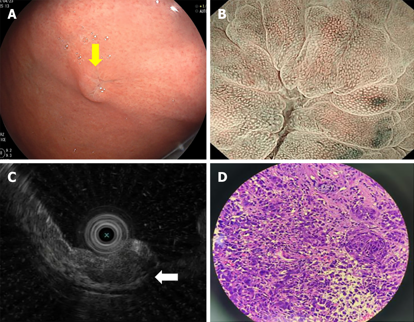
Figure 2 Endoscopy and pathological examinations of the gastric lesion.
A: Gastroscopy showed a 10 mm × 8 mm protruding submucosal tumor with central erosion on the gastric fundus (yellow arrow); B: Magnifying endoscopy showed that the demarcation line of the lesion was not obvious. The microvascular and microsurface patterns were absent on the erosive surface but were still regular on the surrounding mucosa; C: Endoscopic ultrasonography showed a hypoechoic mass with an irregular margin in the submucosal layer, but the submucosal layer had not broken through (white arrow); D: Pathological examination of the gastric biopsy showed poorly differentiated carcinoma (hematoxylin and eosin staining; magnification 100 ×).
- Citation: Lin Y, Wu YL, Zou DD, Luo XL, Zhang SY. Combined gastroscopic and laparoscopic resection of gastric metastatic adenosquamous carcinoma from lung: A case report. World J Gastrointest Surg 2024; 16(9): 3065-3073
- URL: https://www.wjgnet.com/1948-9366/full/v16/i9/3065.htm
- DOI: https://dx.doi.org/10.4240/wjgs.v16.i9.3065


