Copyright
©The Author(s) 2024.
World J Gastrointest Surg. Sep 27, 2024; 16(9): 3065-3073
Published online Sep 27, 2024. doi: 10.4240/wjgs.v16.i9.3065
Published online Sep 27, 2024. doi: 10.4240/wjgs.v16.i9.3065
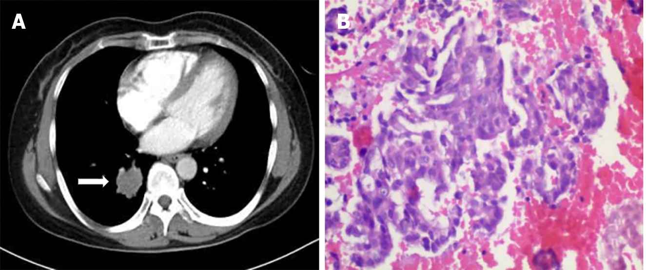
Figure 1 Imaging and pathological examinations of primary lung cancer.
A: Enhanced computed tomography scan of the chest showed a 30 mm × 26 mm tumor in the right lower lobe (white arrow); B: Pathological examination of the lung biopsy showed adenocarcinoma (hematoxylin and eosin staining; magnification 200 ×).
- Citation: Lin Y, Wu YL, Zou DD, Luo XL, Zhang SY. Combined gastroscopic and laparoscopic resection of gastric metastatic adenosquamous carcinoma from lung: A case report. World J Gastrointest Surg 2024; 16(9): 3065-3073
- URL: https://www.wjgnet.com/1948-9366/full/v16/i9/3065.htm
- DOI: https://dx.doi.org/10.4240/wjgs.v16.i9.3065


