Copyright
©The Author(s) 2024.
World J Gastrointest Surg. Sep 27, 2024; 16(9): 3057-3064
Published online Sep 27, 2024. doi: 10.4240/wjgs.v16.i9.3057
Published online Sep 27, 2024. doi: 10.4240/wjgs.v16.i9.3057
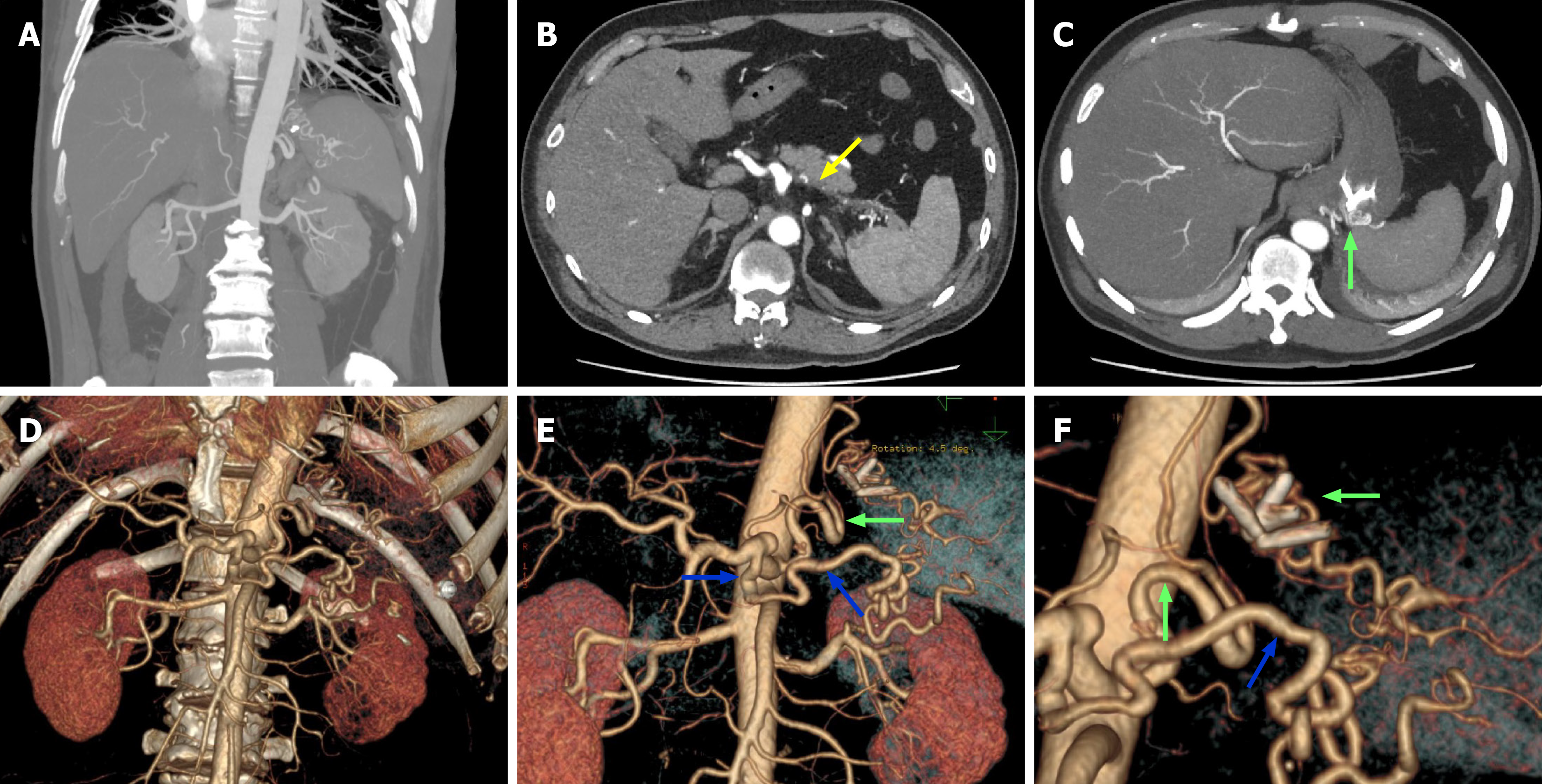
Figure 3 Portal vein computed tomography venography images.
A: Tortuous vessels supplying the spleen observed in the coronal view; B: The splenic artery occlusion indicated by yellow arrows; C: A tortuous artery running through the gastric wall indicated by green arrow; D: A three-dimensional computed tomography image shows the absence of the splenic artery; E and F: The tortuous artery running through the gastric wall (indicated by green arrows), which along with the enlarged dorsal pancreatic artery (indicated by blue arrows), supplies the spleen. The image shows the placement of clips on the gastric wall.
- Citation: Wang H, Tan YQ, Han P, Xu AH, Mu HL, Zhu Z, Ma L, Liu M, Xie HP. Left inferior phrenic arterial malformation mimicking gastric varices: A case report and review of literature. World J Gastrointest Surg 2024; 16(9): 3057-3064
- URL: https://www.wjgnet.com/1948-9366/full/v16/i9/3057.htm
- DOI: https://dx.doi.org/10.4240/wjgs.v16.i9.3057


