Copyright
©The Author(s) 2024.
World J Gastrointest Surg. Sep 27, 2024; 16(9): 2870-2877
Published online Sep 27, 2024. doi: 10.4240/wjgs.v16.i9.2870
Published online Sep 27, 2024. doi: 10.4240/wjgs.v16.i9.2870
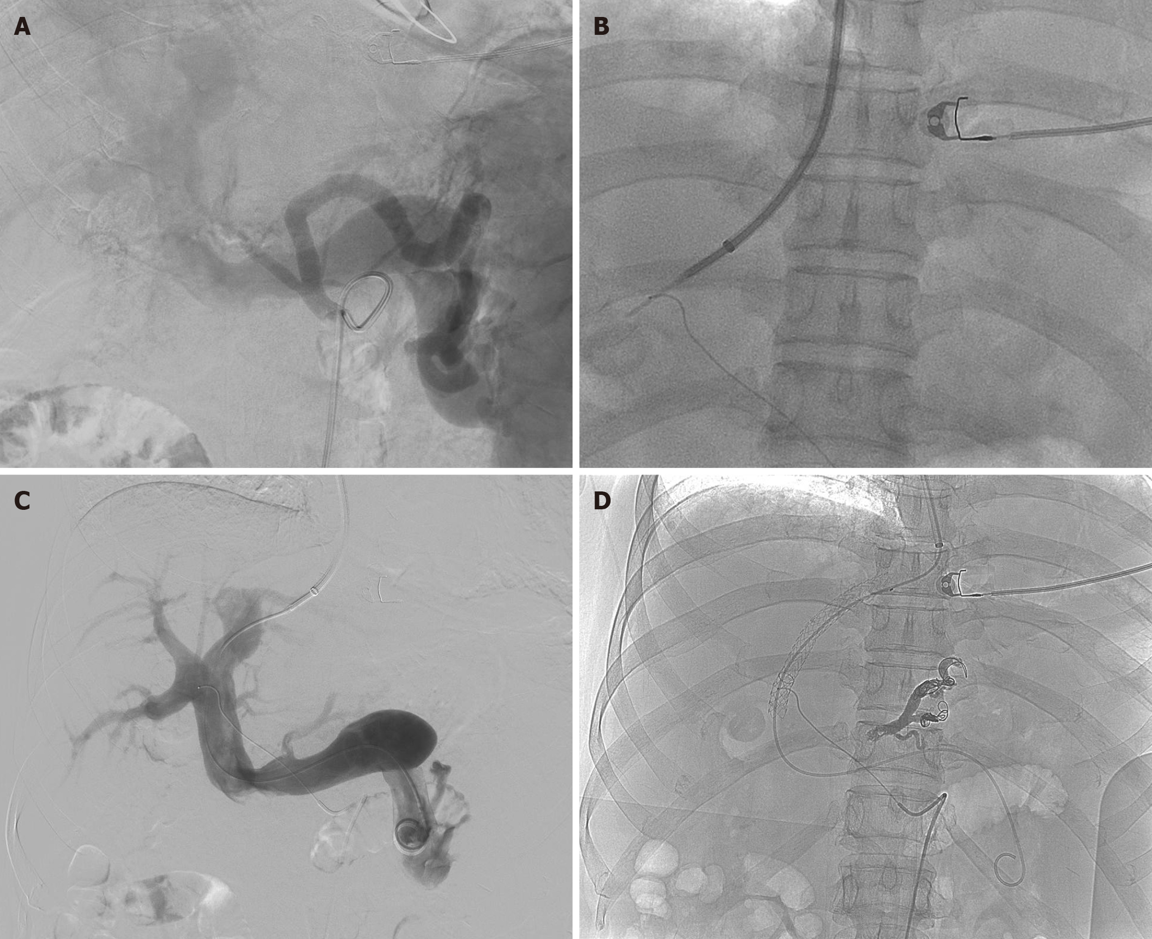
Figure 2 Digital subtraction angiography image overlay technology-assisted hepatic artery labeling for guided transjugular intrahepatic portosystemic shunt procedure.
This figure demonstrates the steps involved in a transjugular intrahepatic portosystemic shunt procedure utilizing digital subtraction angiography (DSA) image overlay technology. A: The simultaneous visualization of the hepatic artery and its adjacent portal vein facilitated by DSA image overlay technology, enabling precise navigation and planning for the procedure; B: The hepatic artery after it has been catheterized and labeled, which serves as a crucial guide for accurate portal vein puncture; C: The portal vein post-catheterization, where angiography confirms that the esophagogastric varices have been fully embolized; D: The final placement of the portosystemic stent with visible spring coils within the embolized esophagogastric varices, indicating successful implementation of the shunt.
- Citation: Li XY, Li Y, Li WQ, Ju S, Dong ZH, Luo JJ. Enhancing transjugular intrahepatic portosystemic shunt procedure efficiency with digital subtraction angiography image overlay technology in esophagogastric variceal bleeding. World J Gastrointest Surg 2024; 16(9): 2870-2877
- URL: https://www.wjgnet.com/1948-9366/full/v16/i9/2870.htm
- DOI: https://dx.doi.org/10.4240/wjgs.v16.i9.2870


