Copyright
©The Author(s) 2024.
World J Gastrointest Surg. Aug 27, 2024; 16(8): 2724-2734
Published online Aug 27, 2024. doi: 10.4240/wjgs.v16.i8.2724
Published online Aug 27, 2024. doi: 10.4240/wjgs.v16.i8.2724
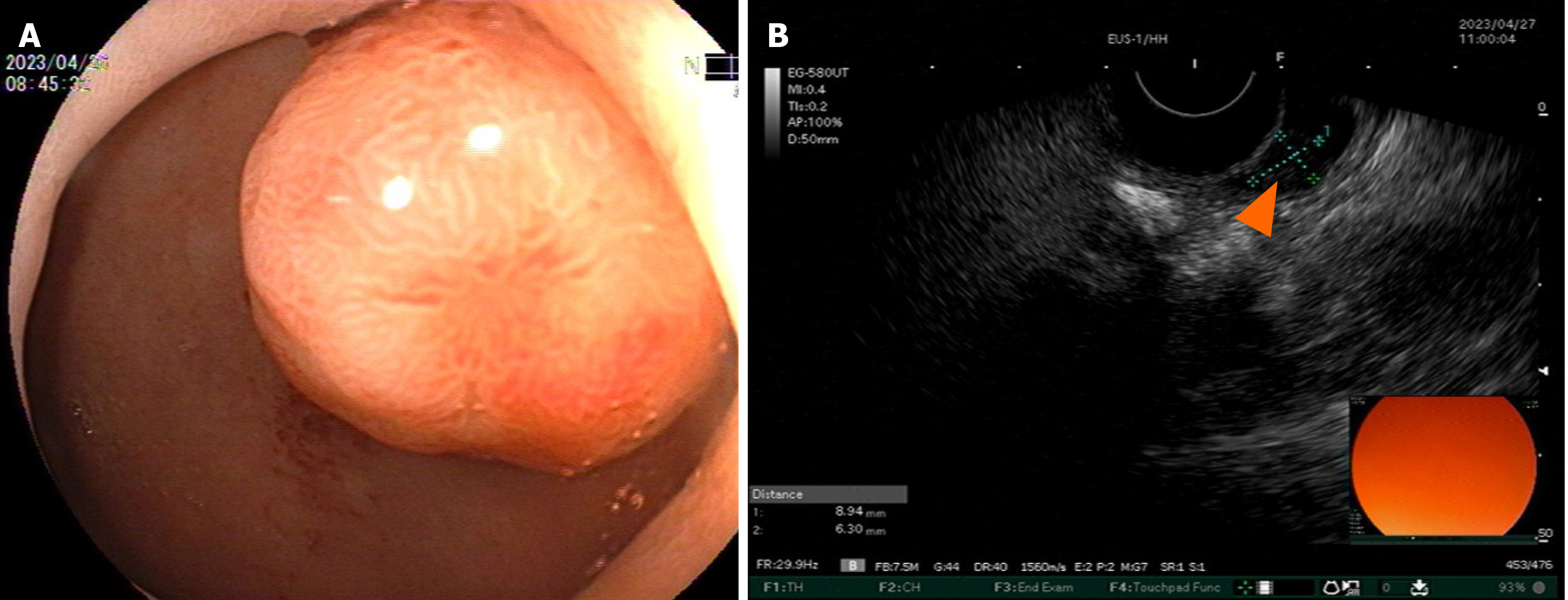
Figure 3 Gastroscopy and endoscopic ultrasonography.
A: Gastroscopy findings of the mass; B: A slightly hypoechoic lesion with a broad base and approximately 8.9 mm × 6.3 mm is seen in the mucosal layer of the anterior wall of the duodenum (orange triangles).
- Citation: Fei S, Wu WD, Zhang HS, Liu SJ, Li D, Jin B. Primary coexisting adenocarcinoma of the colon and neuroendocrine tumor of the duodenum: A case report and review of the literature. World J Gastrointest Surg 2024; 16(8): 2724-2734
- URL: https://www.wjgnet.com/1948-9366/full/v16/i8/2724.htm
- DOI: https://dx.doi.org/10.4240/wjgs.v16.i8.2724


